. 2023 Jul 6;24(13):11134.
doi: 10.3390/ijms241311134.

Effects of Voluntary Physical Exercise on the Neurovascular Unit in a Mouse Model of Alzheimer's Disease

Affiliations

Effects of Voluntary Physical Exercise on the Neurovascular Unit in a Mouse Model of Alzheimer's Disease

Jesús Andrade-Guerrero et al. Int J Mol Sci. .

Abstract

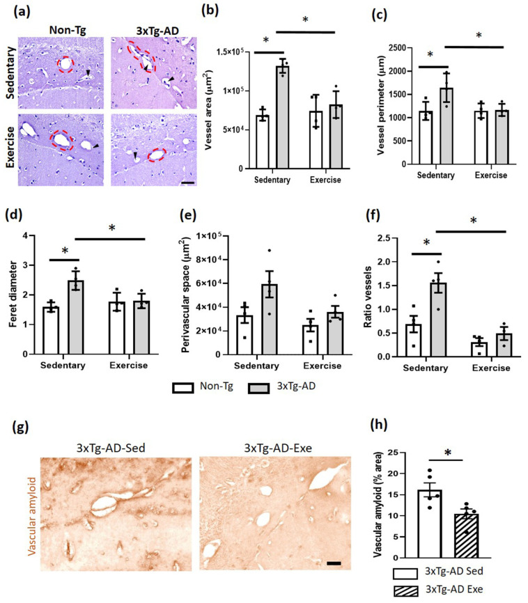
Alzheimer's disease (AD) is the most common neurodegenerative disorder worldwide. Histopathologically, AD presents two pathognomonic hallmarks: (1) neurofibrillary tangles, characterized by intracellular deposits of hyperphosphorylated tau protein, and (2) extracellular amyloid deposits (amyloid plaques) in the brain vasculature (cerebral amyloid angiopathy; CAA). It has been proposed that vascular amyloid deposits could trigger neurovascular unit (NVU) dysfunction in AD. The NVU is composed primarily of astrocytic feet, endothelial cells, pericytes, and basement membrane. Although physical exercise is hypothesized to have beneficial effects against AD, it is unknown whether its positive effects extend to ameliorating CAA and improving the physiology of the NVU. We used the triple transgenic animal model for AD (3xTg-AD) at 13 months old and analyzed through behavioral and histological assays, the effect of voluntary physical exercise on cognitive functions, amyloid angiopathy, and the NVU. Our results show that 3xTg-AD mice develop vascular amyloid deposits which correlate with cognitive deficits and NVU alteration. Interestingly, the physical exercise regimen decreases amyloid angiopathy and correlates with an improvement in cognitive function as well as in the underlying integrity of the NVU components. Physical exercise could represent a key therapeutic approach in cerebral amyloid angiopathy and NVU stability in AD patients.

Keywords: Alzheimer’s disease; astrocytic end-feet; basal membrane; cerebral amyloid angiopathy; cognition; neurovascular unit; pericytes; physical exercise.


Conflict of interest statement

The authors declare no conflict of interest.

Figures

Figure 1
Figure 1
Effects of voluntary physical exercise on anthropometric values and physical performance in 3xTg-AD mice. (a) Body weight was measured over 12 weeks. Repeated measures ANOVA, Tukey’s post hoc, values are means ± SEM (n = 10 per group), * p < 0.05 when compared 3xTg-AD exercise group vs. Non-Tg exercise group. In panel (b), the graph shows gastrocnemius weight after 12 weeks of voluntary physical exercise. Two-ways ANOVA, Tukey’s post hoc, values are means ± SEM (n = 10 per group), * p < 0.05 when compared 3xTg-AD group vs. Non-Tg group. (c) Distance traveled on wheels (km) over 12 weeks. Repeated measures ANOVA, Tukey’s post hoc, values are means ± SEM (n = 10 per group), * p < 0.05. Abbreviations: Exe, exercise; Sed, sedentary.
Figure 2
Figure 2
Effects of voluntary physical exercise on learning and long-term memory via the Barnes Maze. (a) Training latency phase over 4 days. Repeated measures ANOVA, Tukey post hoc, values are means ± SEM (n = 8–9 per group). (b) The graph shows the retention time evaluated 24 h after the training phase. The time in the quadrant (c) and the number of errors (d) are shown in the graphs and representative schemes. Two-way ANOVA, Tukey’s post hoc, values are means ± SEM (n = 8–9 per group), * p < 0.05. Abbreviations: Exe, exercise; Sed, sedentary.
Figure 3
Figure 3
Effect of voluntary physical exercise on Aβ deposition in the hippocampus of the mice. (a) Representative photomicrographs of amyloid deposits of the hippocampal subiculum. Different magnifications (the black dashed box) are illustrated. Scale bar = 500 μm for ×40 magnification; 100 μm for ×100; and 50 μm for ×200. (b) The graph shows the effect of voluntary physical exercise on hippocampal amyloid beta deposits. T student, values are means ± SEM (n = 5 per group), * p < 0.05. Abbreviations: Exe, exercise; Sed, sedentary.
Figure 4
Figure 4
Effects of the voluntary physical exercise on the brain vasculature in the hippocampal fissure of 3xTg-AD mice. (a) Representative photomicrographs of hippocampal fissure blood vessels stained with hematoxylin-eosin under different conditions. The dashed circles show the different vascular morphology between groups and the black arrowheads point to the perivascular space. Scale bar = 20 μm common to all micrographs. The graphs show the area (b), perimeter (c), Feret diameter (d), perivascular space (e), and the ratio of vessels (number of vessels with and without perivascular space; panel (f). Two-way ANOVA, post hoc Tukey, mean ± SEM measured at four anatomical levels (n = 4–5 per group), * p < 0.05. Panel (g) illustrates representative photomicrographs of the vascular amyloid deposits. Scale bar = 20 μm common to all micrographs. (h) The graph shows the measurement of the percentage of area occupied by amyloid vascular deposits. T student, values are mean ± SEM calculated from four anatomical levels (n = 4–5 per group). * p < 0.05. Abbreviations: Exe, exercise; Sed, sedentary.
Figure 5
Figure 5
Effect of voluntary physical exercise on astrocytic end-feet. (a) Representative photomicrographs of AQ4 (astrocytic end-feet) in hippocampal fissure blood vessels. Scale bar = 20 μm common to all micrographs. The black arrow points out the prominent perivascular space. (b) Graphs show astrocyte end-feet area. Two-way ANOVA, Tukey post hoc, mean ± SEM measured at four anatomical levels (n = 5 per group), * p < 0.05. Abbreviations: AQ4, aquaporin-4; Exe, exercise; Sed, sedentary.
Figure 6
Figure 6
Effect of voluntary physical exercise on pericytes and basement membrane in the hippocampus. (a) Fluorescent photomicrographs of pericyte (PDGFR-β; green channel) and basement membrane (collagen IV; red channel) in hippocampal fissure blood vessels. The cellular nuclei were stained with DAPI (blue channel). Scale bar = 20 μm common to all micrographs. The graphs show the area of the pericyte (b) and basement membrane (c). Two-way ANOVA, post hoc Tukey, mean ± SEM measured at four anatomical levels (n = 4–5 per group), * p < 0.05. Abbreviations: Exe, exercise; IF, immunofluorescence; Sed, sedentary.
Figure 7
Figure 7
Correlation between amyloid deposits and neurovascular unit and cognition alteration in sedentary and exercised 3xTg-AD mice. The graphs show the correlation between vascular amyloid deposits and collagen IV (basal membrane) immunofluorescence (IF) area (a), PDGFR-β (pericytes) IF area (b), AQ4 area (astrocytic end-feet) (c), perivascular space (d), and cognition behavior (latency retention time) (e), such as between amyloid plaques and cognition behavior (f), in the hippocampal fissure. Pearson’s correlation coefficient and linear regression appear at the top of every graph p < 0.05 was considered a statistically significant difference.

References

    1. WHO. [(accessed on 10 June 2023)]. Available online: https://www.who.int/es/news-room/fact-sheets/detail/dementia.
    1. Zhang X.X., Tian Y., Wang Z.T., Ma Y.H., Tan L., Yu J.T. The Epidemiology of Alzheimer’s Disease Modifiable Risk Factors and Prevention. J. Prev. Alzheimers Dis. 2021;8:313–321. doi: 10.14283/jpad.2021.15. - DOI - PubMed
    1. Alzheimer’s Association 2021 Alzheimer’s disease facts and figures. Alzheimers Dement. 2021;17:327–406. doi: 10.1002/alz.12328. - DOI - PubMed
    1. Zarahn E., Rakitin B., Abela D., Flynn J., Stern Y. Positive evidence against human hippocampal involvement in working memory maintenance of familiar stimuli. Cereb. Cortex. 2005;15:303–316. doi: 10.1093/cercor/bhh132. - DOI - PubMed
    1. Hyman B.T., Phelps C.H., Beach T.G., Bigio E.H., Cairns N.J., Carrillo M.C., Dickson D.W., Duyckaerts C., Frosch M.P., Masliah E., et al. National Institute on Aging-Alzheimer’s Association guidelines for the neuropathologic assessment of Alzheimer’s disease. Alzheimers Dement. 2012;8:1–13. doi: 10.1016/j.jalz.2011.10.007. - DOI - PMC - PubMed

Substances

Supplementary concepts

LinkOut - more resources