. 2023 Jul 7;24(13):11204.
doi: 10.3390/ijms241311204.

Exploring the Pathophysiologic Cascade Leading to Osteoclastogenic Activation in Gaucher Disease Monocytes Generated via CRISPR/Cas9 Technology

Affiliations

Exploring the Pathophysiologic Cascade Leading to Osteoclastogenic Activation in Gaucher Disease Monocytes Generated via CRISPR/Cas9 Technology

Maximiliano Emanuel Ormazabal et al. Int J Mol Sci. .

Abstract

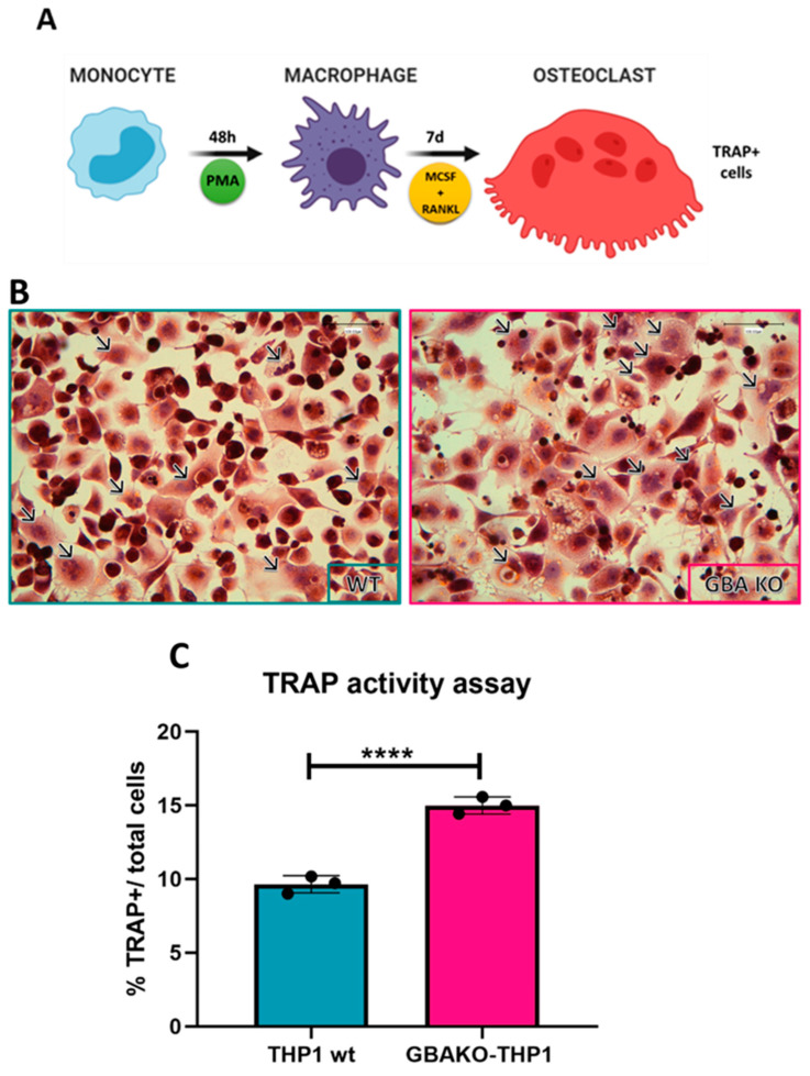
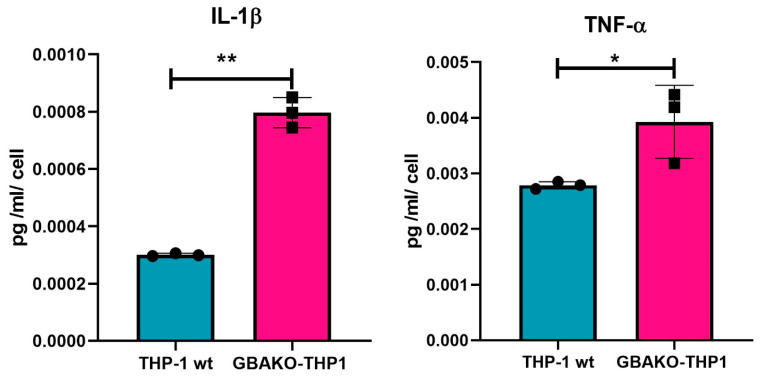
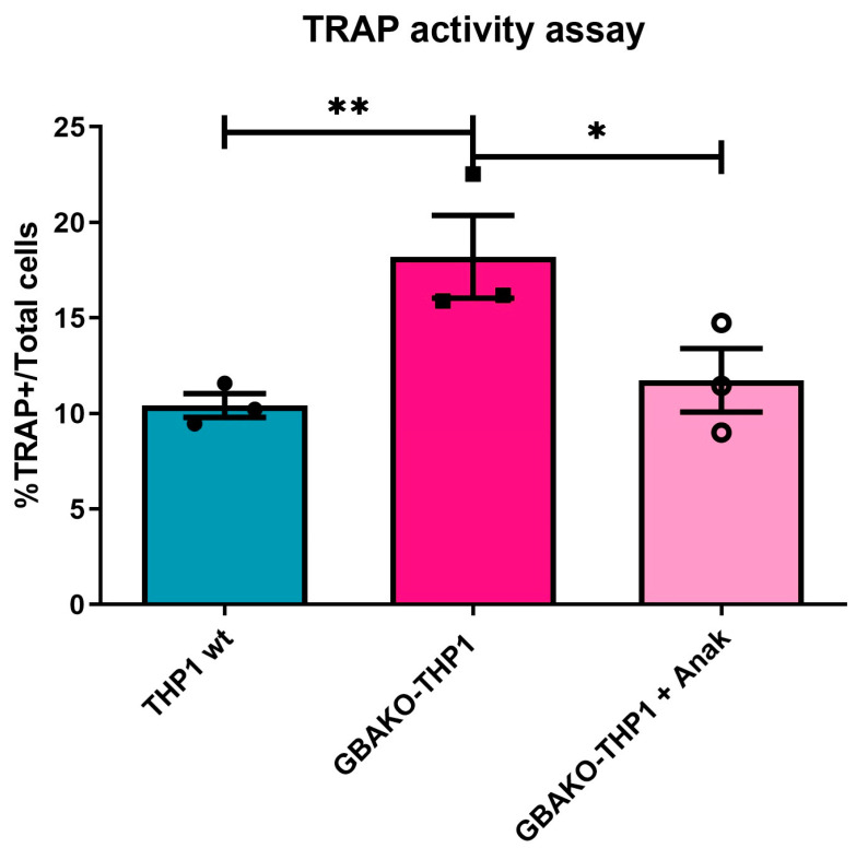
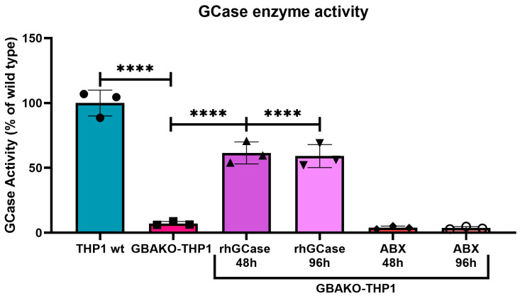
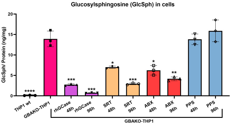
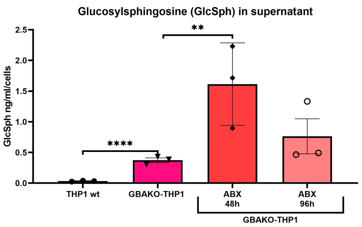
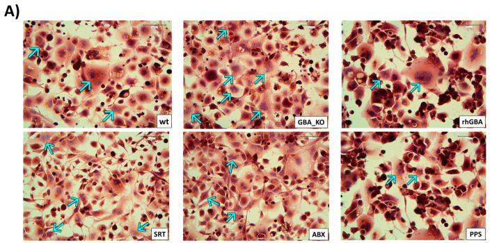
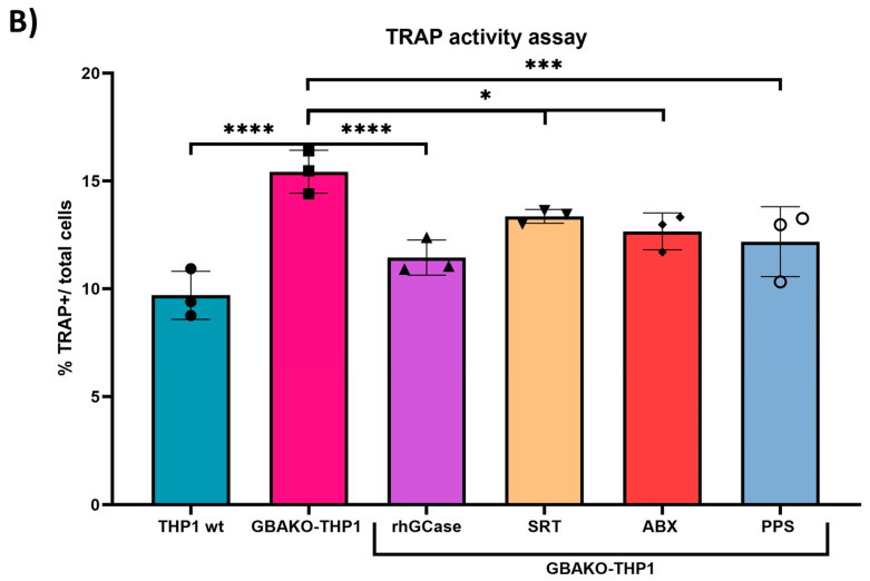
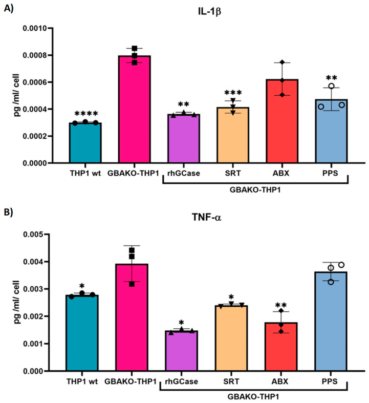
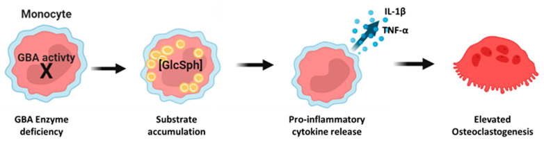
Gaucher disease (GD) is caused by biallelic pathogenic variants in the acid β-glucosidase gene (GBA1), leading to a deficiency in the β-glucocerebrosidase (GCase) enzyme activity resulting in the intracellular accumulation of sphingolipids. Skeletal alterations are one of the most disabling features in GD patients. Although both defective bone formation and increased bone resorption due to osteoblast and osteoclast dysfunction contribute to GD bone pathology, the molecular bases are not fully understood, and bone disease is not completely resolved with currently available specific therapies. For this reason, using editing technology, our group has developed a reliable, isogenic, and easy-to-handle cellular model of GD monocytes (GBAKO-THP1) to facilitate GD pathophysiology studies and high-throughput drug screenings. In this work, we further characterized the model showing an increase in proinflammatory cytokines (Interleukin-1β and Tumor Necrosis Factor-α) release and activation of osteoclastogenesis. Furthermore, our data suggest that GD monocytes would display an increased osteoclastogenic potential, independent of their interaction with the GD microenvironment or other GD cells. Both proinflammatory cytokine production and osteoclastogenesis were restored at least, in part, by treating cells with the recombinant human GCase, a substrate synthase inhibitor, a pharmacological chaperone, and an anti-inflammatory compound. Besides confirming that this model would be suitable to perform high-throughput screening of therapeutic molecules that act via different mechanisms and on different phenotypic features, our data provided insights into the pathogenic cascade, leading to osteoclastogenesis exacerbation and its contribution to bone pathology in GD.

Keywords: Gaucher disease; bone; inflammation; monocytes; osteoclasts.


Conflict of interest statement

The authors declare no conflict of interest.

Figures

Figure 1
Figure 1
Osteoclastogenic differentiation assay in THP-1 wt and GBAKO-THP1 monocytes. (A) Schematic representation of the osteoclast differentiation assay. After inducing macrophage differentiation by incubating cells for 48 h with PMA, cells were cultured in the presence of M-CSF and RANKL to induce osteoclastogenesis. After 7 days, TRAP staining was used to identify the osteoclast-like cells. (B) TRAP staining (brown) and hematoxylin (purple) staining for osteoclasts and nuclei identification, respectively. Arrows indicate osteoclasts (i.e., TRAP+ cells with 3 or more nuclei); scale bar: 100 µm. (C) Quantification of generated osteoclasts expressed as the percentage of the total number of cells that tested TRAP-positive. Data are shown as mean ± SD of three independent experiments. **** p < 0.0001 t-test. Abbreviations: PMA: phorbol-12 myristate-13 acetate; M-CSF: recombinant human macrophage colony-stimulating factor; RANKL: recombinant human sRANK ligand; TRAP: tartrate-resistant acid phosphatase.
Figure 2
Figure 2
IL-1β and TNF-α levels in the culture supernatant of THP-1 wt and GBAKO-THP1 monocytes. The levels of IL-1β and TNF-α released to the culture media of THP-1 wt and GBAKO-THP1 cells were quantified using a Simple Plex assay (ELLA). Data were normalized via the number of cultured cells and expressed as means ± SD of three independent experiments. * p < 0.1 ** p < 0.01 t-test.
Figure 3
Figure 3
Effect of IL-1 β antagonist on the osteoclastogenic potential of GBAKO-THP1 monocytes. GBAKO-THP1 monocytes were differentiated to osteoclasts in the presence or absence of the IL-1 β receptor antagonist Anakinra. The osteoclasts generated were identified as TRAP-positive cells and quantified. The results were expressed as the percentage of the total number of cells that tested TRAP-positive. Data are shown as mean ± SD of three independent experiments. * p < 0.1 ** p < 0.01 t-test.
Figure 4
Figure 4
Effect of treatments on GCase activity in GBAKO-THP1 monocytes. GBAKO-THP1 cells were treated with 1.6 µM of rhGCase or 100 µM of ABX. After 48 or 96 h, the GCase activity was assessed. Only rhGCase treatment resulted in increased GCase activity, while ABX did not show any effect. Data are expressed as the percentage of the GCase activity detected in wild-type cells and are shown as mean ± SD of three independent experiments. **** p < 0.0001 one-way ANOVA.
Figure 5
Figure 5
Effect of different treatments on intracellular GlcSph levels of GBAKO-THP1 monocytes. GBAKO-THP1 cells were treated with rhGCase (1.6 µM), D, L-threo-PDMP (as SRT, 20 µM), ABX (100 µM), and PPS (5 μg/mL) for 48 and 96 h. Intracellular GlcSph was measured using LC-MS/MS. rhGCase, SRT, and ABX treatments significantly reduced intracellular GlcSph levels, but PPS had no effect. Data were normalized by the intracellular protein amount and shown as mean ± SD of three independent experiments. * p < 0.1 ** p < 0.01 *** p < 0.001 **** p < 0.0001 one-way ANOVA.
Figure 6
Figure 6
Effect of Ambroxol on extracellular GlcSph levels of GBAKO-THP1 monocytes. Cells were treated with 100 µM of ABX for 48 and 96 h, and the GlcSph levels in the culture media were measured using LC-MS/MS. Ambroxol treatment increased GlSph release to the culture media. GlcSph levels were normalized by the number of cultured cells, and data are expressed as mean ± SD of three independent experiments. ** p < 0.01 **** p < 0.0001 one-way ANOVA.
Figure 7
Figure 7
Effect of the different treatments on GBAKO-THP1 monocyte osteoclastogenic potential. GBAKO-THP1 cells were treated using rhGCase (1.6 µM), D, L-threo-PDMP (as SRT, 20 µM), ABX (100 µM), and PPS (5 μg/mL) throughout the osteoclastogenic differentiation process. TRAP assay was used to identify the osteoclast-like cells obtained after 7 days. (A) Representative TRAP staining (brown) and hematoxylin (purple) staining for osteoclasts and nuclei identification, respectively. Arrows indicate osteoclasts (i.e., TRAP+ cells with 3 or more nuclei); scale bar: 100 µm. (B) All treatments induced a reduction in the number of osteoclast-like cells differentiated from the GBAKO-THP1 cells. The results are expressed as the percentage of total cells that resulted in TRAP-positive at the end of the differentiation protocol. Data are shown as mean ± SD of three independent experiments. * p < 0.1 *** p < 0.001 **** p < 0.0001 one-way ANOVA.
Figure 7
Figure 7
Effect of the different treatments on GBAKO-THP1 monocyte osteoclastogenic potential. GBAKO-THP1 cells were treated using rhGCase (1.6 µM), D, L-threo-PDMP (as SRT, 20 µM), ABX (100 µM), and PPS (5 μg/mL) throughout the osteoclastogenic differentiation process. TRAP assay was used to identify the osteoclast-like cells obtained after 7 days. (A) Representative TRAP staining (brown) and hematoxylin (purple) staining for osteoclasts and nuclei identification, respectively. Arrows indicate osteoclasts (i.e., TRAP+ cells with 3 or more nuclei); scale bar: 100 µm. (B) All treatments induced a reduction in the number of osteoclast-like cells differentiated from the GBAKO-THP1 cells. The results are expressed as the percentage of total cells that resulted in TRAP-positive at the end of the differentiation protocol. Data are shown as mean ± SD of three independent experiments. * p < 0.1 *** p < 0.001 **** p < 0.0001 one-way ANOVA.
Figure 8
Figure 8
Effect of the different treatments on IL-1β and TNF-α release in GBAKO-THP1 monocytes. GBAKO-THP1 cells were treated with rhGCase (1.6 µM), D, L-threo-PDMP (as SRT, 20 µM), ABX (100 µM), and PPS (5 μg/mL) for 48 h. IL-1β (A) and TNF-α (B) were assessed in the supernatant of cultured cells using Simple Plex assay (ELLA). Results were normalized via the number of cultured cells. Treated GBAKO-THP1 showed decreased levels of one or both cytokines in comparison with untreated GBAKO-THP1. Data are shown as mean ± SD of three independent experiments. * p < 0.1 ** p < 0.01 *** p < 0.001 **** p < 0.0001 t-test.
Figure 9
Figure 9
Schematic pathophysiology in the new monocyte GD model.

References

    1. Stirnemann J.Ô., Belmatoug N., Camou F., Serratrice C., Froissart R., Caillaud C., Levade T., Astudillo L., Serratrice J., Brassier A., et al. A Review of Gaucher Disease Pathophysiology, Clinical Presentation and Treatments. Int. J. Mol. Sci. 2017;18:441. doi: 10.3390/ijms18020441. - DOI - PMC - PubMed
    1. Mistrya P.K., Liua J., Yanga M., Nottolic T., McGratha J., Jaine D., Zhangf K., Keutzerf J., Chuangf W.L., Mehalb W.Z., et al. Glucocerebrosidase Gene-Deficient Mouse Recapitulates Gaucher Disease Displaying Cellular and Molecular Dysregulation beyond the Macrophage. Proc. Natl. Acad. Sci. USA. 2010;107:19473–19478. doi: 10.1073/pnas.1003308107. - DOI - PMC - PubMed
    1. Aflaki E., Moaven N., Borger D.K., Lopez G., Westbroek W., Chae J.J., Marugan J., Patnaik S., Maniwang E., Gonzalez A.N., et al. Lysosomal Storage and Impaired Autophagy Lead to Inflammasome Activation in Gaucher Macrophages. Aging Cell. 2016;15:77–88. doi: 10.1111/acel.12409. - DOI - PMC - PubMed
    1. Bembi B., Marsala S.Z., Sidransky E., Ciana G., Carrozzi M., Zorzon M., Martini C., Gioulis M., Pittis M.G., Capus L. Gaucher’s Disease with Parkinson’s Disease: Clinical and Pathological Aspects. Neurology. 2003;61:99–101. doi: 10.1212/01.WNL.0000072482.70963.D7. - DOI - PubMed
    1. Itzchaki M., Lebel E., Dweck A., Patlas M., Hadas-Halpern I., Zimran A., Elstein D. Orthopedic Considerations in Gaucher Disease since the Advent of Enzyme Replacement Therapy. Acta Orthop. Scand. 2004;75:641–653. doi: 10.1080/00016470410004003. - DOI - PubMed