. 2023 Jun 28;28(13):5047.
doi: 10.3390/molecules28135047.

A Potential Anti-Glioblastoma Compound LH20 Induces Apoptosis and Arrest of Human Glioblastoma Cells via CDK4/6 Inhibition

Affiliations

A Potential Anti-Glioblastoma Compound LH20 Induces Apoptosis and Arrest of Human Glioblastoma Cells via CDK4/6 Inhibition

Yan Wang et al. Molecules. .

Abstract

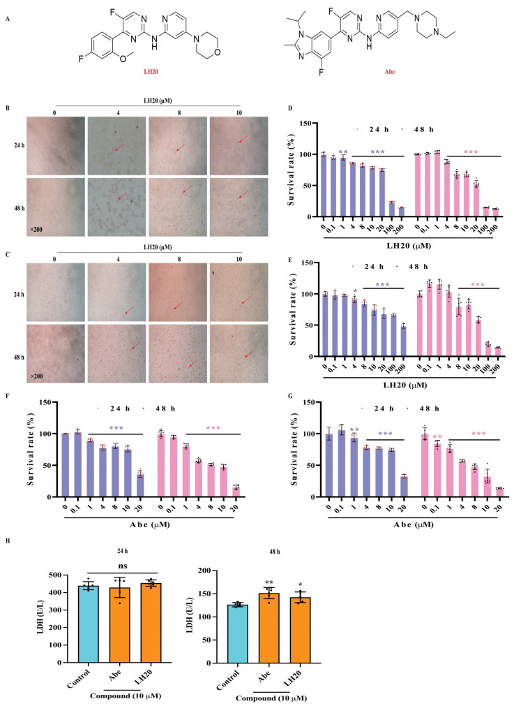
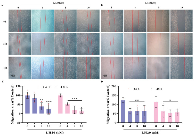
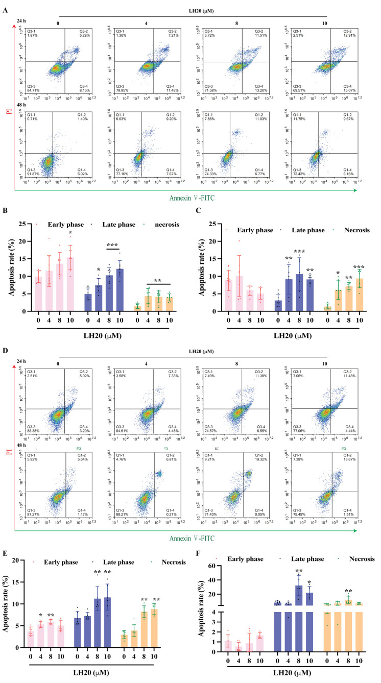
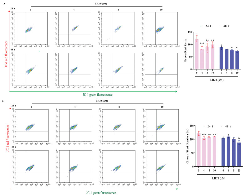
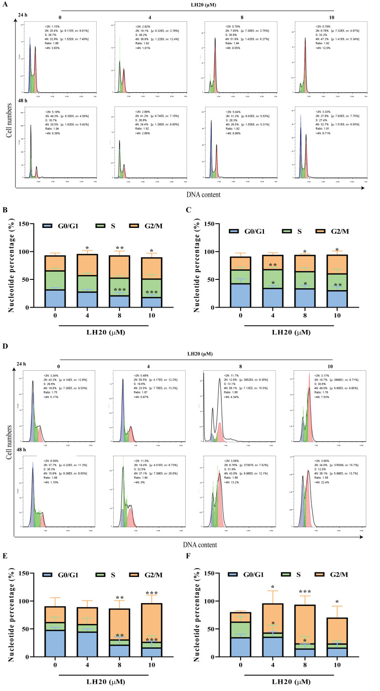
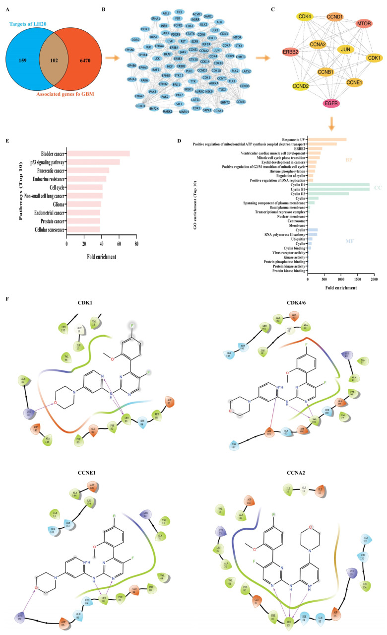
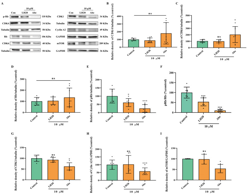
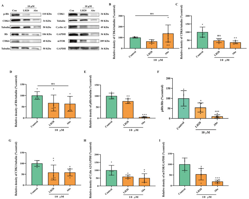
Glioblastoma (GBM) is a deadly brain tumor characterized by signaling dysregulation and aberrant cell cycle control. The CDK4/6-Rb axis is dysregulated in approximately 80% of all GBM cases. In this study, the anti-GBM effect of a novel pyrimidin-2-amine, LH20 was evaluated in vitro using the primary GBM cell lines U87MG and U251. GBM cells were administered LH20 at concentrations of 0.1, 1, 4, 8, 10, 20, 100, and 200 µM for 24 and 48 h, and the proliferation rate was evaluated using a CCK8 assay. Migration, apoptosis, and cell cycle were also assessed using a wound healing assay, Annexin V-FITC/PI apoptosis assay, and cell cycle staining, respectively. The targets of LH20 were predicted using SwissTargetPrediction and molecular docking. Western blotting analysis was performed to confirm the anti-GBM mechanism of LH20. We found that at concentrations of 4, 8, and 10 µM, LH20 significantly inhibited the proliferation and migration of U87MG and U251 cells, induced late phase apoptosis, promoted tumor cell necrosis, and arrested the G2/M phase of the cell cycle. LH20 also inhibited CDK4 and CDK6 activities by decreasing the phosphorylation of Rb. Our results suggest LH20 as a potential treatment strategy against GBM.

Keywords: CDKs; G2/M; LH20; apoptosis; glioblastoma; mitochondrial.


Conflict of interest statement

The authors declare no conflict of interest.

Figures

Figure 1
Figure 1
Effects of LH20 on the survival of GBM cells. (A) Compound structure of LH20 and Abe. (B) Morphological and cell density variation in U87MG cells following treatment for 24 h (top) and 48 h (bottom) with different concentrations of LH20. (C) Morphological and cell density variation in U251 cells observed following treatment for 24 h (top) and 48 h (bottom) with different concentrations of LH20. Red arrows represented the U87MG and U251 cells exhibited morphological changes, including cell rounding and the disappearance of protruding spikes (D) Effects of LH20 on U87MG cell proliferation using a CCK-8 assay (24 h and 48 h). (E) Effects of LH20 on U251 cell proliferation (24 h and 48 h). (F) Effects of Abe on U87MG cell proliferation (24 h and 48 h). (G) Effects of Abe on U251 cell proliferation (24 h and 48 h). (H) Effects of LH20 (10 μM) and Abe (10 μM) on the LDH release in U87MG cells. Data are expressed as mean ± standard deviation; ns represented no significance, * p < 0.05, ** p < 0.01, *** p < 0.001 compared with the untreated or control group.
Figure 2
Figure 2
Effect of LH20 on the migration of U87MG and U251 cells. The migratory potential of GBM cells was analyzed using a wound healing assay. U87MG (A) and U251 (B) cells were incubated in the absence or presence of LH20 (4, 8, and 10 μM) for 48 h. Cells that migrated into the gap were counted under an optical microscope. Red lines indicate the edge of the gap. (C) Migratory area analysis of U87MG cells treated with LH20 for 24 h and 48 h. (D) Migratory area analysis of U251 cells treated with LH20 for 24 h and 48 h. Data are expressed as mean ± standard deviation; * p < 0.05, ** p < 0.01, *** p < 0.001 compared with the untreated group.
Figure 3
Figure 3
LH20 induced apoptosis in GBM cells. (A) Representative graphs from flow cytometry showing U87MG cells. Changes in early apoptosis, late apoptosis, and necrosis rates of U87MG cells treated with LH20 for 24 h (B) and 48 h (C). (D) Representative graphs from flow cytometry showing U251 cells. Changes in early apoptosis, late apoptosis, and necrosis rates of U251 cells treated with LH20 for 24 h (E) and 48 h (F). * p < 0.05, ** p < 0.01, *** p < 0.001 compared with the untreated group.
Figure 4
Figure 4
LH20 induced MMP drop in U87MG (A) and U251 cells (B). * p < 0.05, ** p < 0.01, *** p < 0.001 compared with the untreated group.
Figure 5
Figure 5
LH20 arrested G2/M phase of GBM cells. (A) Representative graphs from flow cytometry showing U87MG cells. Analysis of G0/G1, S, and G2/M phase rate of U87MG cells treated with LH20 for 24 h (B) and 48 h (C). (D) Representative graphs from flow cytometry showing U251 cells. Analysis of G0/G1, S, and G2/M phase rate of U251 cells treated with LH20 for 24 h (E) and 48 h (F). * p < 0.05, ** p < 0.01, *** p < 0.001 compared with the untreated group.
Figure 6
Figure 6
Predicted targets of LH20. (A) Venn graphs showing common genes between GBM and target genes of LH20. (B) Overlapped genes. (C) The 11 genes, nodes with a betweenness of connectivity varied from 129 to 900 were indicated in yellow and pink. (D) Gene ontology (GO) enrichment analysis of the hub genes. Biological process (BP), cellular component (CC), molecular function (MF). (E) Pathway analysis of the hub genes. (F) Molecular docking of LH20 and CDK1, CDK4/6, CCNE1, and CCNA2.
Figure 7
Figure 7
U87MG cells treated with LH20 for 24 h inactivated Rb phosphorylation. (A) Representative blots for CDK4, CDK6, Rb, p-Rb, CDK1, Cyclin A2, and mTOR. (B) Protein expression analysis of CDK4. (C) CDK6. (D) Rb. (E) pRb. (F) Ratio of pRb and Rb. (G) CDK1. (H) Cyclin A2. (I) mTOR. Data are expressed as mean ± standard deviation; ns represented no significance, * p < 0.05, *** p < 0.001 compared with the untreated group.
Figure 8
Figure 8
U87MG cells treated with LH20 for 48 h inactivated Rb phosphorylation. (A) Representative blots for CDK4, CDK6, Rb, p-Rb, CDK1, Cyclin A2, and mTOR. (B) Protein expression analysis of CDK4. (C) CDK6. (D) Rb. (E) pRb. (F) Ratio of pRb and Rb. (G) CDK1. (H) Cyclin A2. (I) mTOR. Data are expressed as mean ± standard deviation; ns represented no significance, * p < 0.05, ** p < 0.01, *** p < 0.001 compared with the untreated group.
Figure 9
Figure 9
NMR spectra of LH20. (A) 1H NMR. (B) 13C NMR.

Similar articles

Cited by

References

    1. Ostrom Q.T., Price M., Neff C., Cioffi G., Waite K.A., Kruchko C., Barnholtz-Sloan J.S. CBTRUS Statistical Report: Primary Brain and Other Central Nervous System Tumors Diagnosed in the United States in 2015–2019. Neuro-Oncol. 2022;24:v1–v95. doi: 10.1093/neuonc/noac202. - DOI - PMC - PubMed
    1. Thakur A., Faujdar C., Sharma R., Sharma S., Malik B., Nepali K., Liou J.P. Glioblastoma: Current Status, Emerging Targets, and Recent Advances. J. Med. Chem. 2022;65:8596–8685. doi: 10.1021/acs.jmedchem.1c01946. - DOI - PMC - PubMed
    1. Fernandes C., Costa A., Osório L., Lago R.C., Linhares P., Carvalho B., Caeiro C. Glioblastoma. Exon Publications; Brisbane City, Australia: 2017. Current Standards of Care in Glioblastoma Therapy; pp. 197–241. - DOI - PubMed
    1. Malumbres M., Barbacid M. Cell cycle, CDKs and cancer: A changing paradigm. Nat. Rev. Cancer. 2009;9:153–166. doi: 10.1038/nrc2602. - DOI - PubMed
    1. Lubanska D., Porter L. Revisiting CDK Inhibitors for Treatment of Glioblastoma Multiforme. Drugs R&D. 2017;17:255–263. doi: 10.1007/s40268-017-0180-1. - DOI - PMC - PubMed

Substances