. 2023 Jun 29;28(13):5088.
doi: 10.3390/molecules28135088.

Hyperoside Nanomicelles Alleviate Atherosclerosis by Modulating the Lipid Profile and Intestinal Flora Structure in High-Fat-Diet-Fed Apolipoprotein-E-Deficient Mice

Affiliations

Hyperoside Nanomicelles Alleviate Atherosclerosis by Modulating the Lipid Profile and Intestinal Flora Structure in High-Fat-Diet-Fed Apolipoprotein-E-Deficient Mice

Yuwen Shi et al. Molecules. .

Abstract

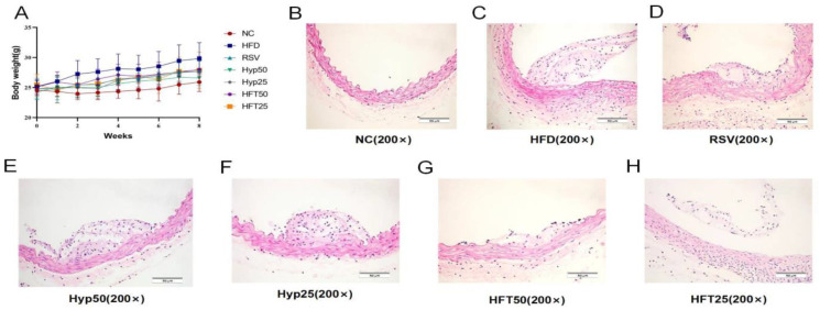
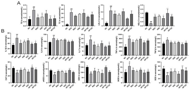
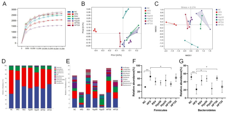
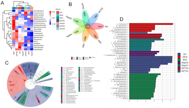
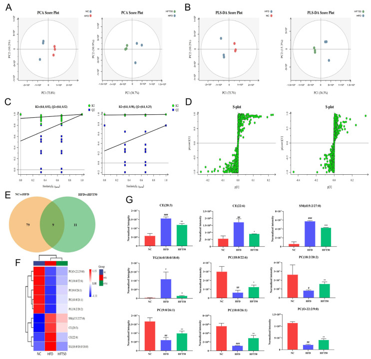
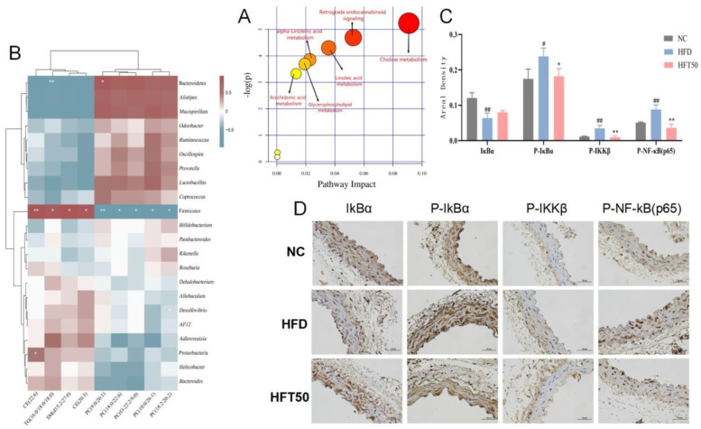
Atherosclerosis (AS) is a serious threat to human health and the main pathological basis of cardiovascular disease. Hyperoside (Hyp), a flavonoid found mainly in traditional Chinese herbs, can exert antitumor, anti-inflammatory, antioxidant, and cardiovascular-protective effects. Herein, we prepared hybrid nanomicelles (HFT) comprising Hyp loaded into pluronic F-127 and polyethylene glycol 1000 vitamin E succinate and assessed their effects on AS. To establish an AS model, apolipoprotein-E-deficient (ApoE-/-) mice were fed a high-fat diet. We then analyzed the effects of HFT on AS-induced changes in aortic tissues and metabolic markers, simultaneously assessing changes in gut flora community structure. In mice with AS, HFT significantly reduced the aortic plaque area; decreased levels of total cholesterol, triglyceride, low-density lipoprotein cholesterol, inflammatory factors, and inducible nitric oxide synthase (NOS); increased high-density lipoprotein cholesterol, endothelial NOS, superoxide dismutase, catalase, and glutathione levels; and promoted the proliferation of beneficial gut bacteria. HFT could regulate intestinal flora structure and lipid metabolism and inhibit inflammatory responses. These beneficial effects may be mediated by inhibiting nuclear factor kappa B signal activation, reducing inflammatory factor expression and improving gut microflora structure and dyslipidemia. The present study provides an empirical basis for the development and clinical application of new dosage forms of Hyp.

Keywords: apolipoprotein E; atherosclerosis; hyperoside nanomicelle; intestinal flora; serum lipidomics.


Conflict of interest statement

The authors declare no conflict of interest.

Figures

Figure 1
Figure 1
Effects of different doses of HFT treatment (eight weeks) on (A) the body weight of ApoE−/− mice (n = 8 per group). (BH) Hematoxylin–eosin staining of atherosclerotic plaque areas at 200× magnification.
Figure 2
Figure 2
Effects of HFT on blood lipid levels in ApoE−/− mice. Variations in (A) TG, TC, LDL-C, and HDL-C content; (B) IL-1β, IL-6, IL-7, TNF-α, TNF-β, CAT, eNOS, GSH, iNOS, and SOD levels (n = 3 per group). ## p < 0.01 and # p < 0.05 compared with the NC group; ** p < 0.01 and * p < 0.05 compared with the HFD group.
Figure 3
Figure 3
HFT affects the alpha and beta diversity and species composition of intestinal flora in ApoE−/− mice. (A) Sparse curves, (B) PCoA, and (C) NMDS. (D) Phylum and (E) genus-level community composition. Relative abundance of (F) Firmicutes and (G) Bacteroidetes at the phylum level (n = 3 per group). * p < 0.05, ** p < 0.01 (correlations).
Figure 4
Figure 4
(A) Heatmap of the top 30 most abundant microbial species at the genus level. (B) Venn diagram, (C) LEfSe, and (D) cladogram. Firmicutes are represented by the red node “b”; Bacteroidetes are represented by the blue node “a”; Chromatiaceae are represented by the dark-red node “b1”; Comamonadaceae are represented by the light-purple node “y”; Clostridia are represented by the purple node “i1”; Alistipes are represented by the dark-green node “f1”; Anaerostipes are represented by the green node “j1.”.
Figure 5
Figure 5
Effects of HFT on lipid metabolites in ApoE−/− mice. (A) PCA score plots, (B) PLS-DA score plots, and (C) displacement test. (D) OPLS-DA permutation plot for the NC vs. HFD (left) and HFD vs. HFT50 groups (right). (E) Venn diagram. (F) Heatmap of nine differentially expressed lipids. (G) Histogram comparing serum differential lipid levels (n = 3 per group). ### p < 0.001, ## p < 0.01, and # p < 0.05 compared with the NC group; *** p < 0.001, ** p < 0.01, and * p < 0.05 compared with the HFD group. HFT, hybrid-mixed nanomicelles comprising Hyp in pluronic F-127 and polyethylene glycol 1000 vitamin E succinate; NC, normal control group; HFD, high-fat diet group; HFT50, HFD + high-dose HFT; PCA, principal component analysis; PLS-DA, a partial least squares-discriminant analysis; OPLS-DA, orthogonal partial least squares-discriminant analysis; PC, phosphatidylcholine; CE, cholesterol ester; SM, sphingomyelin; TG, triglyceride.
Figure 6
Figure 6
(A) Analysis of relevant metabolic pathways. The x-axis represents the topological analysis impact factor, and the y-axis represents the pathway enrichment analysis p-value (−log[P]). (B) Heatmap of serum lipidomics and intestinal flora. (C) Histogram of areal density of IκBα, P-IκBα, P-IKKβ, and P-NF-κB (p65) (n = 3 per group). ## p < 0.01 and # p < 0.05 compared with the NC group, ** p < 0.01 and * p < 0.05 compared with the HFD group. (D) Histological changes in aortic tissues at a magnification of 400×.

References

    1. Ruiz-Leon A.M., Lapuente M., Estruch R., Casas R. Clinical Advances in Immunonutrition and Atherosclerosis: A Review. Front. Immunol. 2019;10:837. doi: 10.3389/fimmu.2019.00837. - DOI - PMC - PubMed
    1. Libby P., Buring J.E., Badimon L., Hansson G.K., Deanfield J., Bittencourt M.S., Tokgozoglu L., Lewis E.F. Atherosclerosis. Nat. Rev. Dis. Prim. 2019;5:56. doi: 10.1038/s41572-019-0106-z. - DOI - PubMed
    1. Voloshyna I., Hussaini S.M., Reiss A.B. Resveratrol in Cholesterol Metabolism and Atherosclerosis. J. Med. Food. 2012;15:763–773. doi: 10.1089/jmf.2012.0025. - DOI - PubMed
    1. Rafieian-Kopaei M., Setorki M., Doudi M., Baradaran A., Nasri H. Atherosclerosis: Process, indicators, risk factors and new hopes. Int. J. Prev. Med. 2014;5:927–946. - PMC - PubMed
    1. Karunakaran D., Nguyen M.A., Geoffrion M., Vreeken D., Lister Z., Cheng H.S., Otte N., Essebier P., Wyatt H., Kandiah J.W., et al. RIPK1 Expression Associates with Inflammation in Early Atherosclerosis in Humans and Can Be Therapeutically Silenced to Reduce NF-κB Activation and Atherogenesis in Mice. Circulation. 2021;143:163–177. doi: 10.1161/CIRCULATIONAHA.118.038379. - DOI - PubMed