. 2023 Jun 29;28(13):5110.
doi: 10.3390/molecules28135110.

An AIE-Active NIR Fluorescent Probe with Good Water Solubility for the Detection of Aβ1-42 Aggregates in Alzheimer's Disease

Affiliations

An AIE-Active NIR Fluorescent Probe with Good Water Solubility for the Detection of Aβ1-42 Aggregates in Alzheimer's Disease

Yan-Ming Ji et al. Molecules. .

Abstract

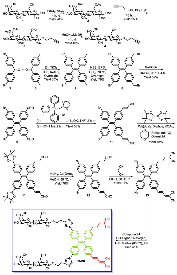
Alzheimer's disease (AD), an amyloid-related disease, seriously endangers the health of elderly individuals. According to current research, its main pathogenic factor is the amyloid protein, which is a kind of fibrillar aggregate formed by noncovalent self-assembly of proteins. Based on the characteristics of aggregation-induced emission (AIE), a bislactosyl-decorated tetraphenylethylene (TPE) molecule TMNL (TPE + malononitrile + lactose), bearing two malononitrile substituents, was designed and synthesized in this work. The amphiphilic TMNL could self-assemble into fluorescent organic nanoparticles (FONs) with near-infrared (NIR) fluorescence emission in physiological PBS (phosphate buffered saline), achieving excellent fluorescent enhancement (47-fold) upon its combination with Aβ1-42 fibrils. TMNL was successfully applied to image Aβ1-42 plaques in the brain tissue of AD transgenic mice, and due to the AIE properties of TMNL, no additional rinsing process was necessary. It is believed that the probe reported in this work should be useful for the sensitive detection and accurate localization mapping of Aβ1-42 aggregates related to Alzheimer's disease.

Keywords: AIE; Alzheimer’s disease; Aβ; amyloid; fluorescence; lactose; near-infrared imaging; tetraphenylethylene.


Conflict of interest statement

The authors declare no conflict of interest.

Figures

Scheme 1
Scheme 1
The synthesis route of TMNL.
Figure 1
Figure 1
The UV absorption spectra and fluorescence emission spectra of TMNL.
Figure 2
Figure 2
(a) The fluorescence spectra of different concentrations (μM) of TMNL in PBS buffer solution (pH 7.4, 10 mM). (b) Scatter plot of fluorescence intensity at 645 nm of different concentrations of TMNL in PBS buffer solution (pH 7.4, 10 mM). λex = 360 nm.
Figure 3
Figure 3
Fluorescence spectra of the excitation (a) and emission (b) of ThT (1 μM)-detecting Aβ1–42 (80 μg·mL−1) fibrils in PBS buffer solution (pH 7.4, 10 mM). λex = 410 nm, λem = 470 nm.
Figure 4
Figure 4
(a) Fluorescence spectra of the interaction of TMNL with Aβ1–42 fibrils after different coincubation times in PBS buffer solution (pH 7.4, 10 mM). Insert: photograph of TMNL after complete interaction with Aβ1–42 fibrils (left) and in the absence of Aβ1–42 fibrils (right) in quartz cuvettes under 365 nm UV light. (b) Scatter plot of the relative fluorescence intensity (I/I0) at 496 nm of the different coincubation times of TMNL and Aβ1–42 fibrils in PBS buffer solution (pH 7.4, 10 mM). I and I0 represent the fluorescence intensity in the presence and absence of Aβ1–42 fibrils, respectively. [TMNL] = 1 μM, [Aβ1–42] = 80 μg·mL−1, λex = 360 nm.
Figure 5
Figure 5
(a) Fluorescence spectra of the interaction of TMNL with different concentrations of Aβ1–42 fibrils (μg·mL−1) in PBS buffer solution (pH 7.4, 10 mM). (b) Scatter plot of the relative fluorescence intensity (I/I0) at 496 nm of the interaction of TMNL and different concentrations of Aβ1–42 fibrils (μg·mL−1) in PBS buffer solution (pH 7.4, 10 mM). I and I0 represent the fluorescence intensity in the presence and absence of Aβ1–42 fibrils, respectively. [TMNL] = 1 μM, λex = 360 nm.
Figure 6
Figure 6
(a) The UV absorption spectra of different concentrations of TMNL in PBS buffer solution (pH 7.4, 10 mM). (b) UV absorption spectra of the interaction of TMNL (1 μM) with different concentrations of Aβ1–42 fibrils (μg·mL−1) in PBS buffer solution (pH 7.4, 10 mM).
Figure 7
Figure 7
(a) Fluorescence spectra of the interaction of TMNL with different interfering substances and Aβ1–42 fibrils in PBS buffer solution (pH 7.4, 10 mM). (b) The histogram of relative fluorescence intensity (I/I0) at 496 nm of the interaction of TMNL with different interfering substances and Aβ1–42 fibrils in PBS buffer solution (pH 7.4, 10 mM). I represents the fluorescence intensity in the presence of the interfering substances or Aβ1–42 fibrils, and I0 represents the fluorescence intensity in the presence of TMNL alone. [Cys] = [GSH] = [HSO3] = [SO32−] = [CN] = 100 μM, [BSA] = [Aβ1–42] = 80 μg·mL−1; [TMNL] = 1 μM; λex = 360 nm.
Figure 8
Figure 8
Saturation binding curves of different concentrations of TMNL interacting with Aβ1–42 fibrils. [Aβ1–42] = 80 μg·mL−1; λex = 360 nm.
Figure 9
Figure 9
(a,b) TEM images of TMNL in PBS buffer solution (pH 7.4, 10 mM). (c,d) TEM images of TMNL after binding to Aβ1–42 fibrils in PBS buffer solution (pH 7.4, 10 mM). [TMNL] = 1 μM; [Aβ1–42] = 80 μg·mL−1.
Figure 10
Figure 10
(a,b) Fluorescence staining images of TMNL in brain tissue sections of AD transgenic mice.

Similar articles

Cited by

References

    1. Gravitz L. Drugs: A tangled web of targets. Nature. 2011;475:S9–S11. doi: 10.1038/475S9a. - DOI - PubMed
    1. Selkoe D.J. Resolving controversies on the path to Alzheimer’s therapeutics. Nat. Med. 2011;17:1060–1065. doi: 10.1038/nm.2460. - DOI - PubMed
    1. Bhasikuttan A.C., Mohanty J. Detection, inhibition and disintegration of amyloid fibrils: The role of optical probes and macrocyclic receptors. Chem. Commun. 2017;53:2789–2809. doi: 10.1039/C6CC08727B. - DOI - PubMed
    1. Hardy J., Selkoe D.J. The Amyloid Hypothesis of Alzheimer’s Disease: Progress and Problems on the Road to Therapeutics. Science. 2002;297:353–356. doi: 10.1126/science.1072994. - DOI - PubMed
    1. Kayed R., Head E., Thompson J.L., McIntire T.M., Milton S.C., Cotman C.W., Glabe C.G. Common Structure of Soluble Amyloid Oligomers Implies Common Mechanism of Pathogenesis. Science. 2003;300:486–489. doi: 10.1126/science.1079469. - DOI - PubMed