. 2023 Jul 2;15(13):2931.
doi: 10.3390/polym15132931.

Chitosan-Collagen Electrospun Nanofibers Loaded with Curcumin as Wound-Healing Patches

Affiliations

Chitosan-Collagen Electrospun Nanofibers Loaded with Curcumin as Wound-Healing Patches

Maila Castellano et al. Polymers (Basel). .

Abstract

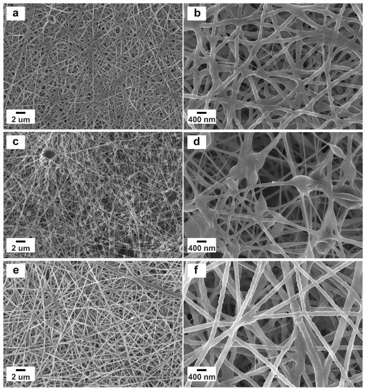
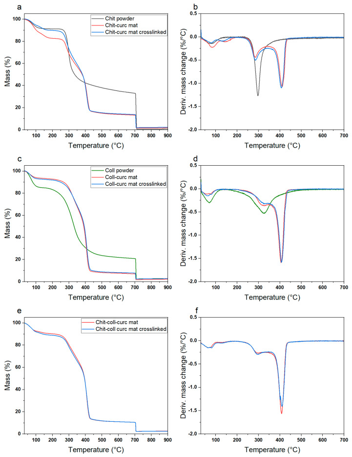
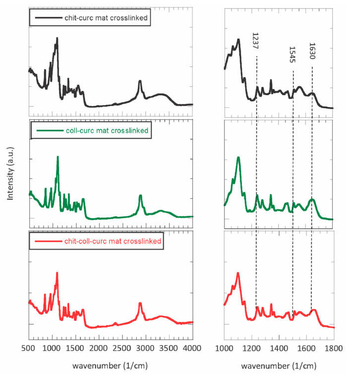
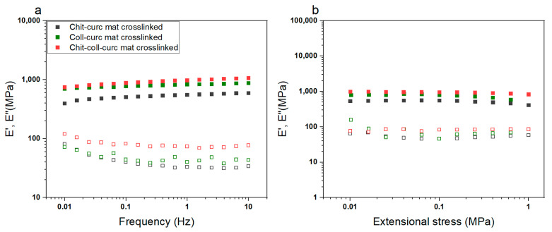
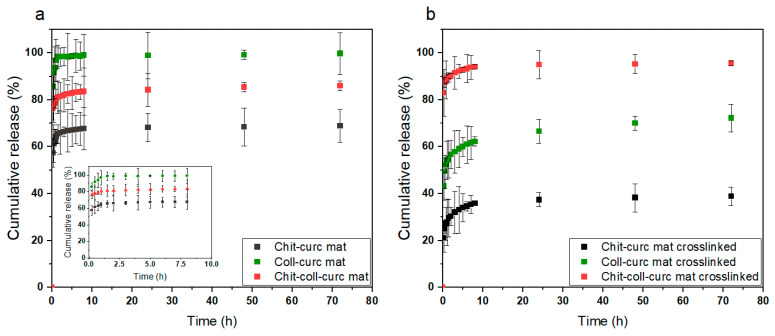
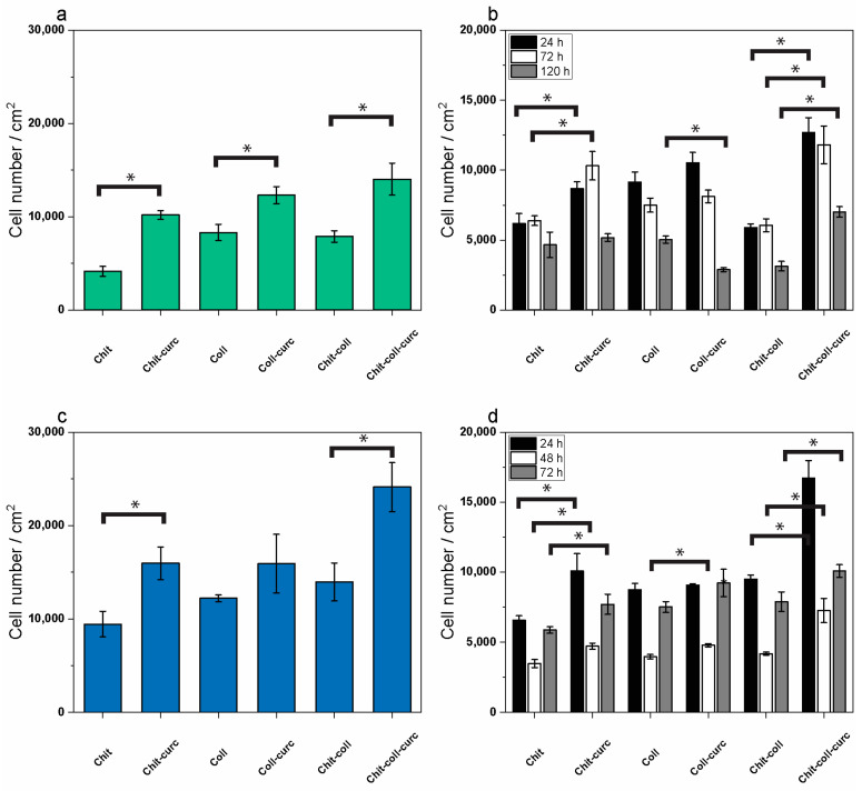
Composite chitosan-collagen nanofibrous mats embedded with curcumin were prepared via a single-step electrospinning procedure and explored as wound-healing patches with superior biological activity. A mild crosslinking protocol consisting of a short exposure to ammonia vapor and UV radiation was developed to ensure proper stability in physiological-like conditions without affecting the intrinsic biocompatibility of chitosan and collagen. The fabricated composite patches displayed a highly porous, homogeneous nanostructure consisting of fibers with an average diameter of 200 nm, thermal stability up to 200 °C, mechanical features able to ensure protection and support to the new tissues, and water-related properties in the ideal range to allow exudate removal and gas exchange. The release kinetic studies carried out in a simulated physiological environment demonstrated that curcumin release was sustained for 72 h when the mats are crosslinked hence providing prolonged bioactivity reflected by the displayed antioxidant properties. Remarkably, combining chitosan and collagen not only ensures prolonged stability and optimal physical-chemical properties but also allows for better-promoting cell adhesion and proliferation and enhanced anti-bacteriostatic capabilities with the addition of curcumin, owing to its beneficial anti-inflammatory effect, ameliorating the attachment and survival/proliferation rates of keratinocytes and fibroblasts to the fabricated patches.

Keywords: chitosan; collagen; curcumin; electrospun nanofibers; wound-healing patches.


Conflict of interest statement

The authors declare no conflict of interest.

Figures

Figure 1
Figure 1
Nanofibrous structure after the crosslinking treatment for (a,b) chitosan–curcumin electrospun mat, (c,d) collagen–curcumin electrospun mat, and (e,f) chitosan–collagen–curcumin electrospun mat. Samples show an overall homogenous morphology, with the composite sample presenting the largest and most regular nanofibers. The absence of observable curcumin crystals indicates their effective encapsulation within the polymeric nanofibers.
Figure 2
Figure 2
TGA and DTGA profiles for (a,b) chitosan-based electrospun mat, (c,d) collagen-based electrospun mat, and (e,f) chitosan–collagen-based electrospun mat. The peaks observed in the DTGA profiles are indicative of the different degradation processes occurring with increasing temperature (i.e., vaporization of the residual humidity at T < 100 °C, degradation of chitosan or collagen at T~300 °C, degradation of PEO at T~400 °C). Notably, the small changes observed between pristine and crosslinked mats indicate the absence of significant physical–chemical changes occurring during the crosslinking treatment.
Figure 3
Figure 3
FTIR spectra for chitosan–curcumin crosslinked mat (grey line), collagen–curcumin crosslinked mat (green line), and chitosan–collagen–curcumin mat (red line). All the absorption bands of the raw materials can be observed along with a slight shift in the position of amide I, II, and III bands of collagen, as highlighted in the enlarged spectra on the right. The observed shifts are likewise due to the occurrence of a new linkage between the polymeric chains.
Figure 4
Figure 4
Dynamic mechanical properties of crosslinked electrospun mats in extensional configuration obtained in (a) frequency sweep mode and (b) stress sweep mode. All samples were within the LVER for the investigated stress range and displayed the typical behavior of solid-like materials with a predominance of the storage modulus over the loss one. Chitosan–collagen–curcumin crosslinked mats were characterized by the highest moduli as a result of the intermolecular interactions between the biopolymers with a consequent increase in the material stiffness.
Figure 5
Figure 5
Curcumin release kinetics in vitro for (a) pristine electrospun mats and (b) crosslinked electrospun mats. Collagen-based mats always presented the highest cumulative release, chitosan-based ones the lowest, and composite patches an intermediate value that agrees with the different stabilities of the samples in physiological conditions. The pristine samples displayed an almost immediate release occurring in the first 5 h, whereas the crosslinked ones were characterized by an initial burst release in the first 10 h followed by a slow release up to 72 h.
Figure 6
Figure 6
Cell adhesion and cell survival/proliferation on membranes as measured by MTT cell viability assays. (a) HaCaT human keratinocyte adhesion to the different membranes with or without curcumin after 16 h incubation. (b) HaCaT human keratinocyte survival/proliferation on the different membranes with or without curcumin after 24 h (black bars), 72 h (white bars), and 120 h (grey bars) of incubation. (c) L929 mouse fibroblast adhesion analyzed in the same conditions as (a). (d) L929 mouse fibroblast proliferation analyzed in the same conditions as (b). Results are expressed as the number of cells per cm2 of each membrane and are the mean ± S.D. of 3 experiments performed in quadruplicate. Statistical analysis is indicated by asterisks. Each asterisk on a bar indicates significance (* p < 0.005) in paired Student t-test between the respective membrane (i.e., collagen, chitosan, or composite) with or without curcumin.

References

    1. Tottoli E.M., Dorati R., Genta I., Chiesa E., Pisani S., Conti B. Skin Wound Healing Process and New Emerging Technologies for Skin Wound Care and Regeneration. Pharmaceutics. 2020;12:735. doi: 10.3390/pharmaceutics12080735. - DOI - PMC - PubMed
    1. Patterson C.W., Stark M., Sharma S., Mundinger G.S. Regeneration and expansion of autologous full-thickness skin through a self-propagating autologous skin graft technology. Clin. Case Rep. 2019;7:2449–2455. doi: 10.1002/ccr3.2533. - DOI - PMC - PubMed
    1. Mastroianni M., Ng Z.Y., Goyal R., Mallard C., Farkash E.A., Leonard D.A., Albritton A., Shanmugarajah K., Kurtz J.M., Sachs D.H., et al. Topical Delivery of Immunosuppression to Prolong Xenogeneic and Allogeneic Split-Thickness Skin Graft Survival. J. Burn Care Res. 2018;39:363–373. doi: 10.1097/BCR.0000000000000597. - DOI - PMC - PubMed
    1. Dong R., Guo B. Smart wound dressings for wound healing. Nano Today. 2021;41:101290. doi: 10.1016/j.nantod.2021.101290. - DOI
    1. Rezvani Ghomi E., Khalili S., Nouri Khorasani S., Esmaeely Neisiany R., Ramakrishna S. Wound dressings: Current advances and future directions. J. Appl. Polym. Sci. 2019;136:47738. doi: 10.1002/app.47738. - DOI

LinkOut - more resources