

Electrospun Poly-L-Lactic Acid Scaffolds Surface-Modified via Reactive Magnetron Sputtering Using Different Mixing Ratios of Nitrogen and Xenon
- PMID: 37447614
- PMCID: PMC10347186
- DOI: 10.3390/polym15132969
Electrospun Poly-L-Lactic Acid Scaffolds Surface-Modified via Reactive Magnetron Sputtering Using Different Mixing Ratios of Nitrogen and Xenon
Abstract
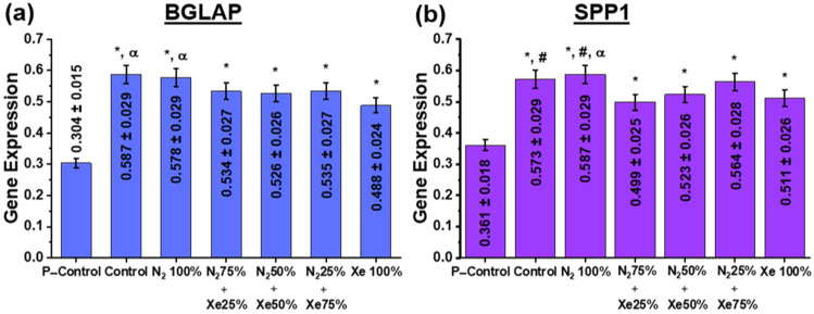
Controlled regeneration processes involving tissue growth using the surface and structure of scaffolds, are actively used in tissue engineering. Reactive magnetron sputtering is a versatile surface modification method of both metal and polymer substrates, as the properties of the formed coatings can be modified in a wide range by changing the process parameters. In magnetron sputtering, the working gas and its composition have an influence on the chemical composition and physical characteristics of the obtained coatings. However, there are no studies addressing the influence of the nitrogen/xenon gas mixture ratio in direct current magnetron sputtering on the deposition rate, physicochemical and in vitro properties of surface-modified biocompatible poly-L-lactic acid scaffolds. In this study, the application of mixtures of nitrogen and xenon in various ratios is demonstrated to modify the surface of non-woven poly-L-lactic acid scaffolds by direct current magnetron sputtering of a titanium target. It has been found that the magnetron sputtering parameters chosen do not negatively influence the morphology of the prepared scaffolds, but increase the hydrophilicity. Moreover, quantitative spectroscopic analysis results indicate that the formed coatings are primarily composed of titanium oxide and titanium oxynitride compounds and is dependent on the gas mixture ratio only to a certain extent. Atomic force microscopy investigations of the roughness of the fibers of the electrospun scaffolds and the thickness of the coatings formed on them show that the considerable variations observed in the intrinsic fiber reliefs are due to the formation of a fine layer on the fiber surfaces. The observed decrease in roughness after plasma modification is due to temperature and radiation effects of the plasma. In vitro experiments with human osteosarcoma cells show that the scaffolds investigated here have no cytotoxic effect on these cells. The cells adhere and proliferate well on each of the surface-modified electrospun scaffolds, with stimulation of cell differentiation in the osteogenic direction.
Keywords: HOS cells; electrospun PLLA scaffold; nitrogen-containing titanium coating; proliferative activity; reactive magnetron sputtering; working gas mixtures.
Conflict of interest statement
The authors declare no conflict of interest.
Figures


Similar articles
-
Surface modification of electrospun poly-(l-lactic) acid scaffolds by reactive magnetron sputtering.Colloids Surf B Biointerfaces. 2018 Feb 1;162:43-51. doi: 10.1016/j.colsurfb.2017.11.028. Epub 2017 Nov 12. Colloids Surf B Biointerfaces. 2018. PMID: 29149727
-
Surface Modification of Electrospun Bioresorbable and Biostable Scaffolds by Pulsed DC Magnetron Sputtering of Titanium for Gingival Tissue Regeneration.Polymers (Basel). 2022 Nov 15;14(22):4922. doi: 10.3390/polym14224922. Polymers (Basel). 2022. PMID: 36433049 Free PMC article.
-
Antibacterial Activity and Cytocompatibility of Electrospun PLGA Scaffolds Surface-Modified by Pulsed DC Magnetron Co-Sputtering of Copper and Titanium.Pharmaceutics. 2023 Mar 14;15(3):939. doi: 10.3390/pharmaceutics15030939. Pharmaceutics. 2023. PMID: 36986800 Free PMC article.
-
Ion-substituted calcium phosphate coatings by physical vapor deposition magnetron sputtering for biomedical applications: A review.Acta Biomater. 2019 Apr 15;89:14-32. doi: 10.1016/j.actbio.2019.03.006. Epub 2019 Mar 6. Acta Biomater. 2019. PMID: 30851454 Review.
-
Comparison of CrN Coatings Prepared Using High-Power Impulse Magnetron Sputtering and Direct Current Magnetron Sputtering.Materials (Basel). 2023 Sep 20;16(18):6303. doi: 10.3390/ma16186303. Materials (Basel). 2023. PMID: 37763579 Free PMC article. Review.
Cited by
-
Three-Dimensional Cross-Linking Network Coating for the Flame Retardant of Bio-Based Polyamide 56 Fabric by Weak Bonds.Polymers (Basel). 2024 Apr 10;16(8):1044. doi: 10.3390/polym16081044. Polymers (Basel). 2024. PMID: 38674963 Free PMC article.
References
-
- Abbasi N., Hamlet S., Love R.M., Nguyen N.-T. Porous scaffolds for bone regeneration. J. Sci. Adv. Mater. Devices. 2020;5:1–9. doi: 10.1016/j.jsamd.2020.01.007. - DOI
-
- Agarwal S. Biodegradable Polymers: Present Opportunities and Challenges in Providing a Microplastic-Free Environment. Macromol. Chem. Phys. 2020;221:2000017. doi: 10.1002/macp.202000017. - DOI
-
- Ibrahim H.M., Klingner A. A review on electrospun polymeric nanofibers: Production parameters and potential applications. Polym. Test. 2020;90:106647. doi: 10.1016/j.polymertesting.2020.106647. - DOI
Grants and funding
LinkOut - more resources
Full Text Sources
