

Comparative Evaluations on Real-Time Monitoring of Temperature Sensors during Endoscopic Laser Application
- PMID: 37447918
- PMCID: PMC10346888
- DOI: 10.3390/s23136069
Comparative Evaluations on Real-Time Monitoring of Temperature Sensors during Endoscopic Laser Application
Abstract
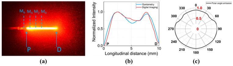
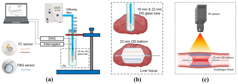
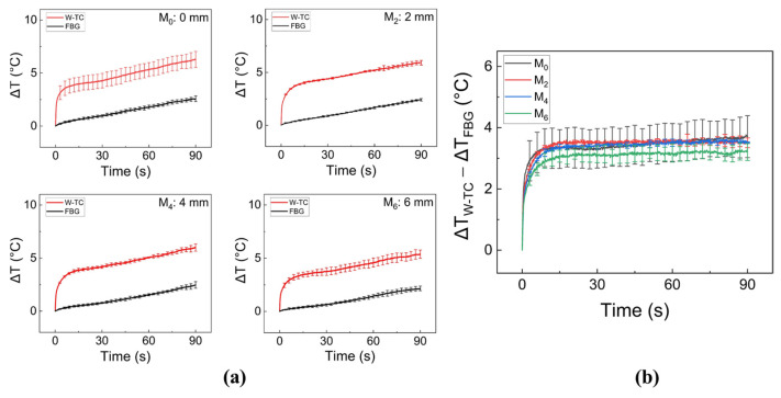
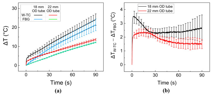
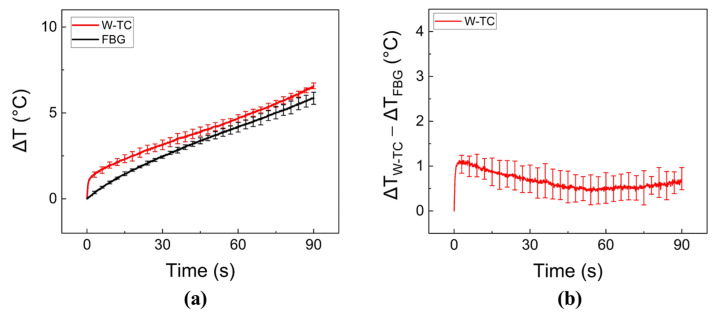
Temperature sensors, such as Fiber Bragg Grating (FBG) and thermocouple (TC), have been widely used for monitoring the interstitial tissue temperature during laser irradiation. The aim of the current study was to compare the performance of both FBG and TC in real-time temperature monitoring during endoscopic and circumferential laser treatment on tubular tissue structure. A 600-µm core-diameter diffusing applicator was employed to deliver 980-nm laser light (30 W for 90 s) circumferentially for quantitative evaluation. The tip of the TC was covered with a white tube (W-TC) in order to prevent direct light absorption and to minimize temperature overestimation. The temperature measurements in air demonstrated that the measurement difference in the temperature elevations was around 3.5 °C between FBG and W-TC. Ex vivo porcine liver tests confirmed that the measurement difference became lower (less than 1 °C). Ex vivo porcine esophageal tissue using a balloon-integrated catheter exhibited that both FBG and W-TC consistently showed a comparable trend of temperature measurements during laser irradiation (~2 °C). The current study demonstrated that the white tube-covered TC could be a feasible sensor to monitor interstitial tissue temperature with minimal overestimation during endoscopic laser irradiation. Further in vivo studies on gastroesophageal reflux disease will investigate the performance of the W-TC to monitor the temperature of the esophageal mucosa surface in real-time mode to warrant the safety of endoscopic laser treatment.
Keywords: Fiber Bragg Grating; diffusing fiber; thermal coagulation; thermocouple.
Conflict of interest statement
The authors declare that there is no conflict of interest regarding the publication of this paper. V.G.T. is the employee and H.W.K. is the founder and CEO of TeCure, Inc. TeCure, Inc. had no role in the decision to publish the results.
Figures

Similar articles
-
Real-time temperature monitoring with fiber Bragg grating sensor during diffuser-assisted laser-induced interstitial thermotherapy.J Biomed Opt. 2017 Apr 1;22(4):45008. doi: 10.1117/1.JBO.22.4.045008. J Biomed Opt. 2017. PMID: 28425558
-
Development of Novel Balloon-Integrated Optical Catheter for Endoscopic and Circumferential Laser Application.Ann Biomed Eng. 2023 Sep;51(9):2021-2034. doi: 10.1007/s10439-023-03228-8. Epub 2023 May 16. Ann Biomed Eng. 2023. PMID: 37191825
-
In situ temperature measurements with thermocouple probes during laser interstitial thermotherapy (LITT): quantification and correction of a measurement artifact.Lasers Surg Med. 1998;23(2):94-103. doi: 10.1002/(sici)1096-9101(1998)23:2<94::aid-lsm7>3.0.co;2-q. Lasers Surg Med. 1998. PMID: 9738544
-
Feasibility of fiber Bragg grating and long-period fiber grating sensors under different environmental conditions.Sensors (Basel). 2010;10(11):10105-27. doi: 10.3390/s101110105. Epub 2010 Nov 10. Sensors (Basel). 2010. PMID: 22163460 Free PMC article.
-
Feasibility study of endoscopic thermal coagulation with circumferential laser irradiation for treating esophageal tissue.Lasers Med Sci. 2020 Jun;35(4):893-900. doi: 10.1007/s10103-019-02877-3. Epub 2019 Oct 22. Lasers Med Sci. 2020. PMID: 31641966
Cited by
-
Quantitative investigations on light emission profiles for interstitial laser treatment.Biomed Opt Express. 2024 Nov 20;15(12):6877-6892. doi: 10.1364/BOE.540470. eCollection 2024 Dec 1. Biomed Opt Express. 2024. PMID: 39679393 Free PMC article.
References
-
- Köksoy F.N., Gönüllü D. The Benign Strictures of the Esophagus. JAREM J. Acad. Res. Med. 2016;6:1–14. doi: 10.5152/jarem.2015.777. - DOI
-
- Murat Ferhat F., Taner K. Anatomy of Esophagus. In: Chai J., editor. Esophageal Abnormalities. IntechOpen; Rijeka, Croatia: 2017. Chapter 1.
MeSH terms
Grants and funding
LinkOut - more resources
Full Text Sources
