

Assessment of Morphological Variations of the Coronoid Process, Condyle, and Sigmoid Notch as an Adjunct in Personal Identification Using Orthopantomograms Among the North Indian Population
- PMID: 37448437
- PMCID: PMC10336369
- DOI: 10.7759/cureus.40275
Assessment of Morphological Variations of the Coronoid Process, Condyle, and Sigmoid Notch as an Adjunct in Personal Identification Using Orthopantomograms Among the North Indian Population
Abstract
Aim: The aim of this study is to assess morphological variations of the coronoid process, condyle, and sigmoid notch as an adjunct in personal identification using orthopantomograms among the North Indian population.
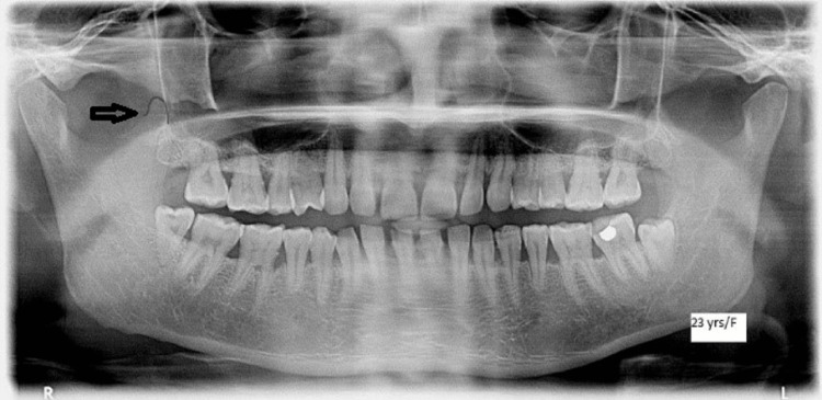
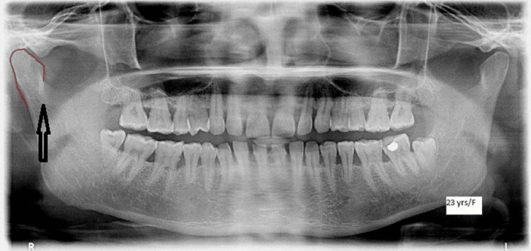
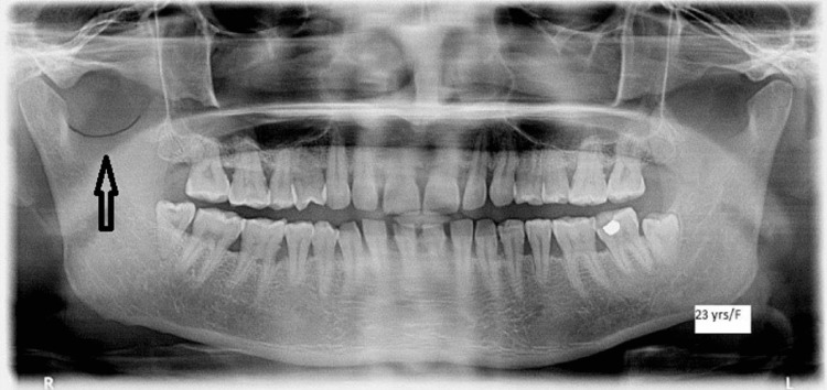
Methodology: The study sample (n=240) was distributed into four age groups: Group I: 30 males and 30 females aged 10-19 years, Group II: 30 males and 30 females aged 20-29 years, Group III: 30 males and 30 females aged 30-39 years, and Group IV: 30 males and 30 females aged 40-59 years. All were subjected to panoramic radiographs. The different morphological forms of the coronoid process, condyle, and sigmoid notch were evaluated.
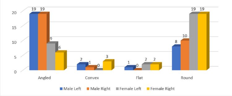
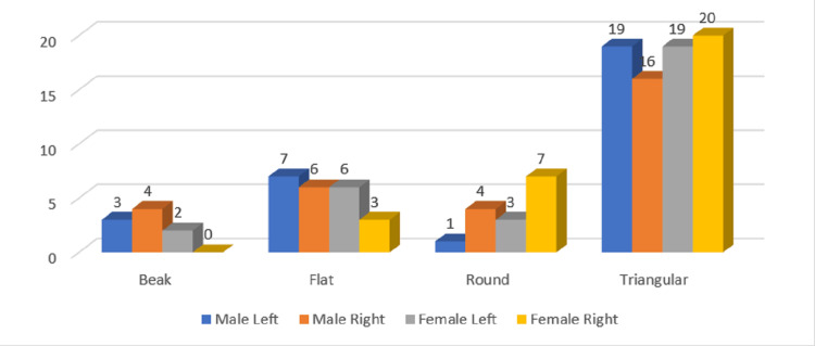
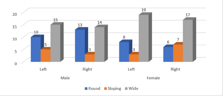
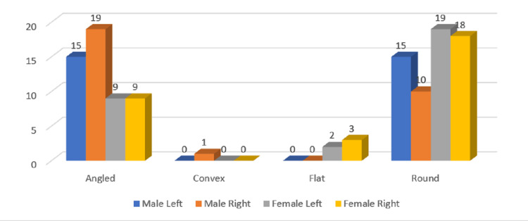
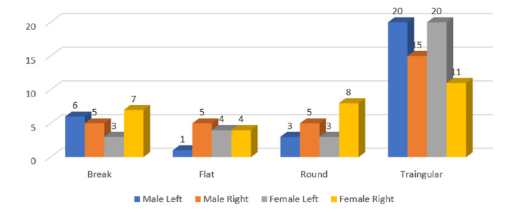
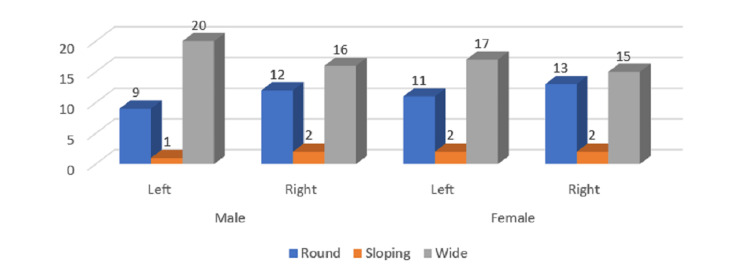
Results: The results showed that across all age groups, angular condyles were the most common kind of condyle in males, followed by round and convex types. The present study found that the coronoid process typically takes on a triangle shape across all ages and sexes. Additionally, the vast majority of cases were triangular on both sides, and this was true across both sexes. It was found in this study that the sigmoid notch most commonly took the form of a larger notch, followed by a rounder notch.
Conclusion: Using panoramic photos to portray the different morphologies of the coronoid process, condyle, and sigmoid notch can be a much simpler and faster method of identifying an individual, especially in the event of a mass disaster, so long as antemortem data are kept. The method of radiographic identification of individuals has recently gained prominence due to its efficacy. Radiographs like these can be invaluable in forensic dentistry, where they can help unearth previously hidden evidence if premortem records are retained. As a potential approach for individual identification among our population, panoramic radiographs were used to investigate the varying morphological forms of the coronoid process, condyle, and sigmoid notch.
Keywords: condyle; coronoid process; orthopantomograms; panoramic radiograph; sigmoid notch.
Copyright © 2023, Bains et al.
Conflict of interest statement
The authors have declared that no competing interests exist.
Figures
References
-
- The shape of the condyle and position of the meniscus in temporomandibular joint dysfunction. Juniper RP. Br J Oral Maxillofac Surg. 1994;32:71–76. - PubMed
-
- Morphologic changes in the temporomandibular joint in different age groups: an autopsy investigation. Pereira FJ Jr, Lundh H, Westesson PL. Oral Surg Oral Med Oral Pathol. 1994;78:279–287. - PubMed
-
- Impact of TMJ radiographs on clinician decision making. White SC, Pullinger AG. Oral Surg Oral Med Oral Pathol Oral Radiol Endod. 1995;79:375–381. - PubMed
-
- Condylar appearance in panoramic radiograms of asymptomatic subjects and patients with temporomandibular disorders. Honda E, Yoshino N, Sasaki T. Oral Radiol. 1994;10:43–53.
-
- Morphology of coronoid process and sigmoid notch in orthopantomograms of South Indian population. Shakya S, Ongole R, Nagraj SK. World J Dent. 2013;4:1–3.
LinkOut - more resources
Full Text Sources
Research Materials