Review
. 2022 Oct 5;5(1):20220011.
doi: 10.1259/bjro.20220011. eCollection 2023.

Pictorial guide for variants of Covid-19: CT imaging and interpretation

Affiliations
Review

Pictorial guide for variants of Covid-19: CT imaging and interpretation

Giacomo Bonito et al. BJR Open. .

Abstract

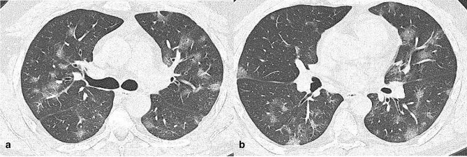
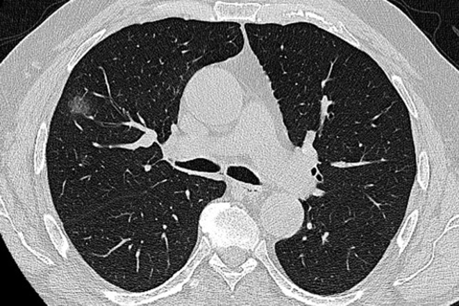
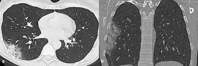
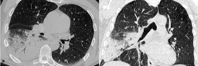
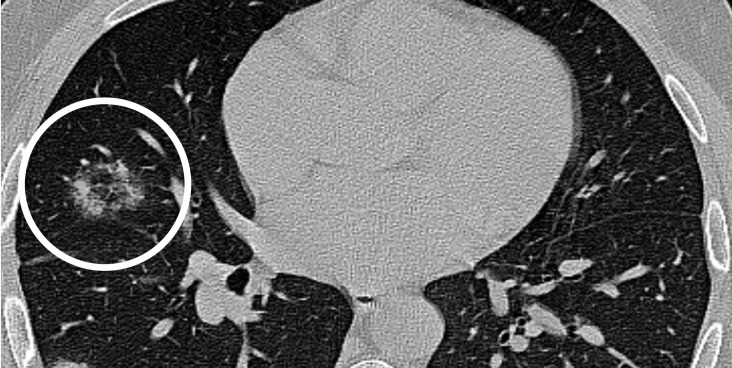
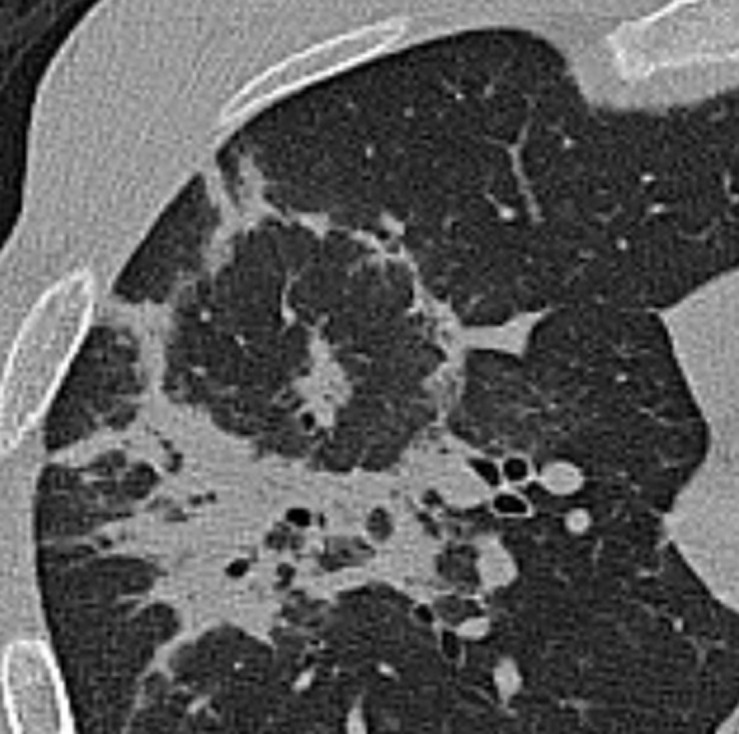
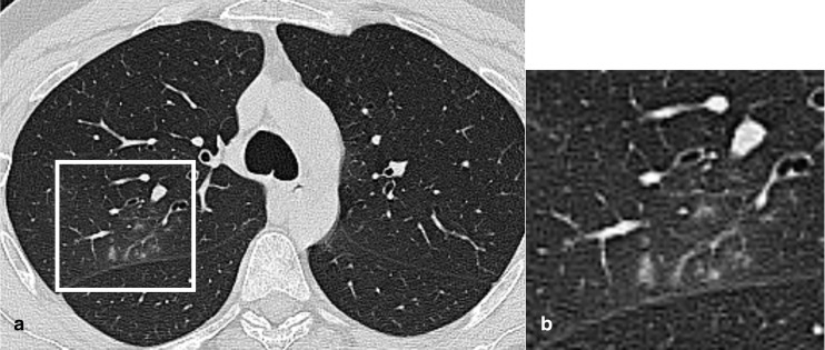
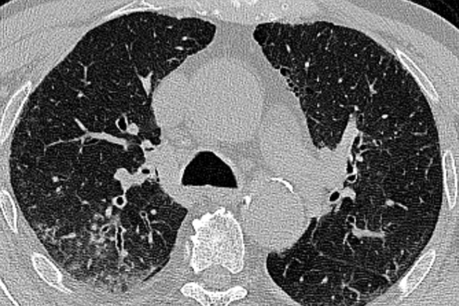
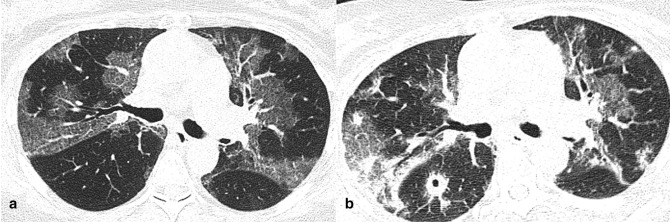
Typical radiologic images of Covid-19 pneumonia consists in a wide spectrum of chest manifestations, which range from peripheral predominant ground-glass opacities to an organizing pneumonia pattern, with additional features including crazy-paving, consolidations, fibrotic streaks and linear opacities. With variants imaging profile of Covid-19 evolves, producing relatively atypical/indeterminate CT pattern of pulmonary involvement, which overlap with imaging features of a variety of other respiratory diseases, including infections, drug reaction and hypersensitivity pneumonia. Our knowledge of these radiological findings is incomplete and there is a need to strengthen the recognition of the many faces of Covid-19 pneumonia.


Figures

Figure 1.
Figure 1.
Axial CT images (a, b) of a 34-year-old male infected by SARS-CoV-2 α variant (3 days since symptoms onset). CT showed centrally and peripherally located multifocal GGO opacities with mainly involvement of upper lobes.
Figure 2.
Figure 2.
A 54-year-old female COVID −19 patient (2 days since symptoms onset) infected by Omicron variant. CT scan shows single ground glass opacity with rounded appearance, in the right upper lobe.
Figure 3.
Figure 3.
A 78-year-old male arriving at the emergency department with fever. Axial (a) and coronal reformatted (b) CT images documented a subpleural GGO in right lower lobe. Notably, there were signs of consolidation in the context of the lesion. The real-time reverse transcription polymerase chain reaction test was positive for Sars-Cov-2 infection. Subsequent genetic sequencing detected α variant. GGO, ground-glass opacity.
Figure 4.
Figure 4.
A 57-year-old COVID-19 patient (δ variant) presenting fever and dry cough for 3 days. Axial (a) and coronal (b) reformatted CT images showed pneumonia with isolated lobar consolidation in the right lower lobe.
Figure 5.
Figure 5.
A 32-year-old female infected with COVID-19 (δ Variant): axial unenhanced CT image showed focus of central ground glass nodule surrounded by an inner ring of air and an outer ring of ground glass (white circle). This appearance ressembles a bullseye.
Figure 6.
Figure 6.
The “bullseye sign” in a 44-year-old male COVID-19 patient (Omicron variant) with fever and dry cough for 7 days.
Figure 7.
Figure 7.
Axial CT image of a 25-year-old male Covid-19 patient (γ variant) showed perifissural small centrilobular nodules connected to linear branching opacities (white box), in right upper lobe.
Figure 8.
Figure 8.
Axial CT image of a 78-year-old male Covid-19 patient (α variant), showed tree-in-bud opacities in right upper lobe with bronchial wall thickening.
Figure 9.
Figure 9.
CT images of a 58-year-old male patient with confirmed COVID-19 pneumonia (γ variant). Axial (a) and coronal reformatted (b) images showed diffuse lung involvement with ill-defined centrilobular GGO and subpleural sparing. GGO, ground-glass opacity.
Figure 10.
Figure 10.
A 42-year-old female COVID-19 patient (γ variant) presenting fever and chills for 4 days. Axial (a) and coronal reformatted (b) images showed bilateral ill-defined centrilobular GGO. GGO, ground-glass opacity.
Figure 11.
Figure 11.
Axial CT image of a 68-year-old patient with COVID-19 pneumonia (δ variant), with symptoms for 4 days, showed bilateral extensive consolidations. The patient died 5 days later.
Figure 12.
Figure 12.
Axial CT image slices of a female COVID-19 patient (α Variant) of 81-year-old. a) CT scan, performed after 1 day from symptoms onset, showed a mixed pattern of GGO and consolidation in upper lobes with peribronchovascular involvement. b) CT obtained after 5 days from symtoms onset. Lesions increased with higher density and superimposed thickening of the interlobular septum in both lungs, with predominant dorsal consolidations.
Figure 13.
Figure 13.
Evolution of CT findings in a 56-year-old female COVID-19 patient (δ variant). (a) CT scan, performed after 3 days from symptom onset, showed GGO with superimposed inter- and intralobular septal thickening (crazy-paving pattern) in upper lobes. (b) Scan obtained on day 7 from symptom onset, showed peripheral consolidations. In apical segment of right lower lobe, CT demonstrated rounded consolidation with cavitation in context.
Figure 14.
Figure 14.
A 53-year-old female COVID-19 patient (Omicron variant), presenting with dry cough, dyspnea and fever up to 39° for 3 days. Axial CT image showed large amount of pericardial effusion.

Similar articles

References

    1. Kanne JP, Bai H, Bernheim A, Chung M, Haramati LB, Kallmes DF, et al. . COVID-19 imaging: what we know now and what remains unknown. Radiology 2021; 299: E262–79. doi: 10.1148/radiol.2021204522 - DOI - PMC - PubMed
    1. Boehm E, Kronig I, Neher RA, Eckerle I, Vetter P, Kaiser L, et al. . Novel SARS-cov-2 variants: the pandemics within the pandemic. Clin Microbiol Infect 2021; 27: 1109–17. doi: 10.1016/j.cmi.2021.05.022 - DOI - PMC - PubMed
    1. Kumar S, Thambiraja TS, Karuppanan K, Subramaniam G. Omicron and delta variant of sars-cov-2: A comparative computational study of spike protein. J Med Virol 2022; 94: 1641–49. doi: 10.1002/jmv.27526 - DOI - PubMed
    1. Hochhegger B, Zanon M, Altmayer S, Mandelli NS, Stüker G, Mohammed T-L, et al. . COVID-19 mimics on chest CT: a pictorial review and radiologic guide. Br J Radiol 2021; 94(1118): 20200703. doi: 10.1259/bjr.20200703 - DOI - PMC - PubMed
    1. Ye Z, Zhang Y, Wang Y, Huang Z, Song B. Chest CT manifestations of new coronavirus disease 2019 (COVID-19): a pictorial review. Eur Radiol 2020; 30: 4381–89. doi: 10.1007/s00330-020-06801-0 - DOI - PMC - PubMed