

Role of neonatal cerebrospinal fluid cytology in correlation to C-reactive protein, blood culture, risk factors and clinical outcomes in neonatal intensive care
- PMID: 37448924
- PMCID: PMC10336944
- DOI: 10.4103/jfmpc.jfmpc_980_21
Role of neonatal cerebrospinal fluid cytology in correlation to C-reactive protein, blood culture, risk factors and clinical outcomes in neonatal intensive care
Abstract
Introduction: The number of neonatal cerebrospinal fluid (CSF) samples sent from the neonatal intensive care unit (NICU) for cytologic examination is rising, warranting accurate analysis and interpretation of the same. This study was taken up to assess the usefulness of CSF cell count and cytology in NICU settings, as it can be used even in a resource-limited setting.
Aim and objective: 1) To study the prevalence of cell count and cytologic changes in CSF from NICU and assess their usefulness in correlation to C-reactive protein, CSF neutrophil percentage, blood, CSF culture, and other biochemical parameters. 2) To correlate cell counts and cytology with age, period of gestation, presence, and absence of sepsis, seizures, intracranial hemorrhage, and their clinical follow-up.
Materials and methods: A retrospective study was done on neonatal CSF samples submitted for cytology over one year (January-December 2016) in the Department of Pathology. CSF cell counts were retrieved, and cytosmears were reviewed for cellularity, cell type, proportion, and background and correlated with the biochemical, microbiological, and clinicoradiological findings.
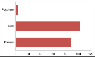
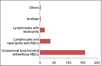
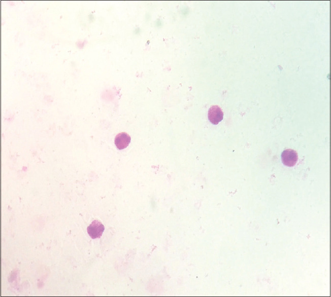
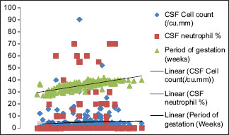
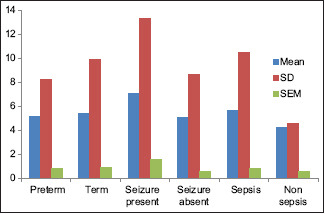
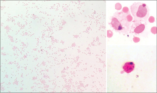
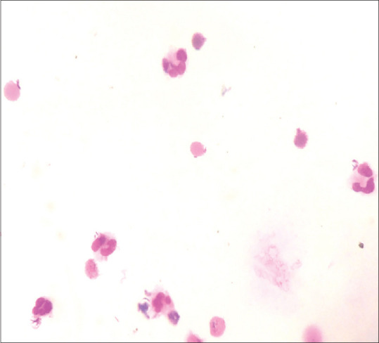
Results: A total of 213 samples were included with 140 males and 73 females with an age range of 0-28 (mean: 7.3) days. The mean CSF cell count was 5.48/cu.mm (0-90 cells/cu.mm). The most frequent cytologic finding was occasional lymphocytes or acellular CSF (63.9%). The CSF leucocyte count and protein levels showed a significant correlation with s C-reactive protein. The CSF cytology showed a significant correlation between the age of the neonate and blood neutrophil percentage (P = 0.0158). History of intracranial hemorrhage showed a significantly higher frequency of the presence of red blood cells (P = 0.0147).
Conclusion: Accurate cell counts, cytology of neonatal CSF, and biochemical and microbiological workup can help diagnose and manage neonates in intensive care.
Keywords: Hemorrhage; meningitis; preterm.
Copyright: © 2023 Journal of Family Medicine and Primary Care.
Conflict of interest statement
There are no conflicts of interest.
Figures
Similar articles
-
Reference range for cerebrospinal fluid values in neonates: 5-year retrospective study.J Matern Fetal Neonatal Med. 2022 Dec;35(26):10584-10590. doi: 10.1080/14767058.2022.2139172. Epub 2022 Oct 30. J Matern Fetal Neonatal Med. 2022. PMID: 36310086
-
Neonatal meningitis: what is the correlation among cerebrospinal fluid cultures, blood cultures, and cerebrospinal fluid parameters?Pediatrics. 2006 Apr;117(4):1094-100. doi: 10.1542/peds.2005-1132. Pediatrics. 2006. PMID: 16585303
-
Diagnostic utility of combined immature and total neutrophil counts along with C-reactive protein in early detection of neonatal sepsis: A cross-sectional study.Ann Med Surg (Lond). 2022 Apr 9;77:103589. doi: 10.1016/j.amsu.2022.103589. eCollection 2022 May. Ann Med Surg (Lond). 2022. PMID: 35637988 Free PMC article.
-
Overview of cerebrospinal fluid cytology.Handb Clin Neurol. 2017;145:563-571. doi: 10.1016/B978-0-12-802395-2.00035-3. Handb Clin Neurol. 2017. PMID: 28987194 Review.
-
Normal Values for Cerebrospinal Fluid in Neonates: A Systematic Review.Neonatology. 2021;118(6):629-638. doi: 10.1159/000517630. Epub 2021 Oct 5. Neonatology. 2021. PMID: 34818234
Cited by
-
Does C-reactive protein help to diagnose the infection in new-born and neonatal in context to maternal immunological marker? An opinion.J Family Med Prim Care. 2023 Nov;12(11):3005-3006. doi: 10.4103/jfmpc.jfmpc_993_23. Epub 2023 Nov 21. J Family Med Prim Care. 2023. PMID: 38186789 Free PMC article. No abstract available.
-
Neonatal cerebrospinal fluid cytology: Preanalytical and analytical phase considerations.J Family Med Prim Care. 2024 Mar;13(3):1134-1135. doi: 10.4103/jfmpc.jfmpc_1519_23. Epub 2024 Apr 4. J Family Med Prim Care. 2024. PMID: 38736836 Free PMC article. No abstract available.
References
-
- Rahimi J, Woehrer A. Overview of cerebrospinal fluid cytology. Handb Clin Neurol. 2017;145:563–571. 4. - PubMed
-
- Celik IH, Demirel G, Canpolat FE, Dilmen U. A common problem for neonatal intensive care units:Late preterm infants, a prospective study with term controls in a large perinatal center. J Matern Fetal Neonatal Med. 2013;26:459–62. - PubMed
-
- Glatstein MM, Zucker-Toledano M, Arik A, Scolnik D, Oren A, Reif S. Incidence of traumatic lumbar puncture:Experience of a large, tertiary care pediatric hospital. Clin Pediatr (Phila) 2011;50:1005–9. - PubMed
LinkOut - more resources
Full Text Sources
Research Materials