Review
. 2023 Jun 28:16:1207007.
doi: 10.3389/fnmol.2023.1207007. eCollection 2023.

Remyelination in animal models of multiple sclerosis: finding the elusive grail of regeneration

Affiliations
Review

Remyelination in animal models of multiple sclerosis: finding the elusive grail of regeneration

Davin Packer et al. Front Mol Neurosci. .

Abstract

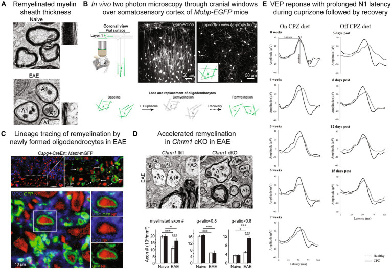
Remyelination biology and the therapeutic potential of restoring myelin sheaths to prevent neurodegeneration and disability in multiple sclerosis (MS) has made considerable gains over the past decade with many regeneration strategies undergoing tested in MS clinical trials. Animal models used to investigate oligodendroglial responses and regeneration of myelin vary considerably in the mechanism of demyelination, involvement of inflammatory cells, neurodegeneration and capacity for remyelination. The investigation of remyelination in the context of aging and an inflammatory environment are of considerable interest for the potential translation to progressive multiple sclerosis. Here we review how remyelination is assessed in mouse models of demyelination, differences and advantages of these models, therapeutic strategies that have emerged and current pro-remyelination clinical trials.

Keywords: OPC; demyelination; multiple sclerosis; oligodendrocyte; remyelination.


Conflict of interest statement

The authors declare that the research was conducted in the absence of any commercial or financial relationships that could be construed as a potential conflict of interest.

Figures

Figure 1
Figure 1
Remyelination assessment in mouse models. (A) Myelin sheath visualization by electron microscopy with thinner myelin sheaths in remyelinated axons (A1) in EAE compared to naïve spinal cord. (B) In vivo two photon microscopy through cranial windows over the somatosensory cortex of Mobp-EGFP mice allows for visualization of loss and replacement of oligodendrocytes and myelin sheaths. (C) Lineage tracing approach to visualize myelin sheaths from newly born oligodendrocytes utilizing Cspg4-CreER™; Mapt-mGFP mice and tamoxifen injection prior to EAE induction. New myelin sheaths generated by OPCs express mGFP reporter (green) and wrap neurofilament positive axons (red). (D) Electron microscopy of conditional knockout of muscarinic acetylcholine receptor Chrm1 in oligodendrocytes (Chrm1 cKO: Cnp-Cre+; Chrm1fl/fl) in EAE results in reduced axonal loss and enhanced remyelination in spinal cord indicated by significantly increased axons with g-ratios >0.8. Black bars indicate Chrm1 cKO and white bars Cnp-Cre-; Chrm1fl/fl control. A1-remyelinated axon, A2-demyelinated axon. (E) Longitudinal visual evoked potential (VEP) waveforms during and off cuprizone (CPZ) diet. N1 latency is prolonged on cuprizone diet and recovers after return to normal diet. Black lines-healthy/normal diet mice, gray lines-cuprizone treated mice. (A,C,D) Reproduced from Mei et al. (2016a), accelerated remyelination during inflammatory demyelination prevents axonal loss and improves functional recovery, eLife © Creative Commons. (B) Reproduced from Orthmann-Murphy et al. (2020), remyelination alters the pattern of myelin in the cerebral cortex, eLife © Creative Commons. (E) Reproduced from Marenna et al. (2022), visual evoked potentials to monitor myelin cuprizone-induced functional changes, Frontiers in Neuroscience © Creative Commons.

Similar articles

Cited by

References

    1. Absinta M., Maric D., Gharagozloo M., Garton T., Smith M. D., Jin J., et al. . (2021). A lymphocyte-microglia-astrocyte axis in chronic active multiple sclerosis. Nature 597, 709–714. doi: 10.1038/s41586-021-03892-7 - DOI - PMC - PubMed
    1. Antel J. P., Lin Y. H., Cui Q. L., Pernin F., Kennedy T. E., Ludwin S. K., et al. . (2019). Immunology of oligodendrocyte precursor cells in vivo and in vitro. J. Neuroimmunol. 331, 28–35. doi: 10.1016/j.jneuroim.2018.03.006 - DOI - PubMed
    1. Arnett H. A., Mason J., Marino M., Suzuki K., Matsushima G. K., Ting J. P. Y. (2001). TNF alpha promotes proliferation of oligodendrocyte progenitors and remyelination. Nat. Neurosci. 4, 1116–1122. doi: 10.1038/nn738 - DOI - PubMed
    1. Babbe H., Roers A., Waisman A., Lassmann H., Goebels N., Hohlfeld R., et al. . (2000). Clonal expansions of CD8(+) T cells dominate the T cell infiltrate in active multiple sclerosis lesions as shown by micromanipulation and single cell polymerase chain reaction. J. Exp. Med. 192, 393–404. doi: 10.1084/jem.192.3.393 - DOI - PMC - PubMed
    1. Bacmeister C. M., Barr H. J., McClain C. R., Thornton M. A., Nettles D., Welle C. G., et al. . (2020). Motor learning promotes remyelination via new and surviving oligodendrocytes. Nat. Neurosci. 23, 819–831. doi: 10.1038/s41593-020-0637-3 - DOI - PMC - PubMed