. 2023 Jun 21:4:100084.
doi: 10.1016/j.nbas.2023.100084. eCollection 2023.

Upregulation of endocytic protein expression in the Alzheimer's disease male human brain

Affiliations

Upregulation of endocytic protein expression in the Alzheimer's disease male human brain

Mouhamed Alsaqati et al. Aging Brain. .

Abstract

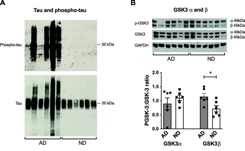
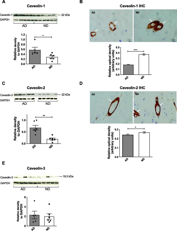
Amyloid-beta (Aβ) is produced from amyloid precursor protein (APP) primarily after APP is internalised by endocytosis and clathrin-mediated endocytic processes are altered in Alzheimer's disease (AD). There is also evidence that cholesterol and flotillin affect APP endocytosis. We hypothesised that endocytic protein expression would be altered in the brains of people with AD compared to non-diseased subjects which could be linked to increased Aβ generation. We compared protein expression in frontal cortex samples from men with AD compared to age-matched, non-diseased controls. Soluble and insoluble Aβ40 and Aβ42, the soluble Aβ42/Aβ40 ratio, βCTF, BACE1, presenilin-1 and the ratio of phosphorylated:total GSK3β were significantly increased while the insoluble Aβ42:Aβ40 ratio was significantly decreased in AD brains. Total and phosphorylated tau were markedly increased in AD brains. Significant increases in clathrin, AP2, PICALM isoform 4, Rab-5 and caveolin-1 and 2 were seen in AD brains but BIN1 was decreased. However, using immunohistochemistry, caveolin-1 and 2 were decreased. The results obtained here suggest an overall increase in endocytosis in the AD brain, explaining, at least in part, the increased production of Aβ during AD.

Keywords: Alzheimer’s disease; Amyloid precursor protein; Amyloid-beta; Endocytosis; Human brain; Male sex.


Conflict of interest statement

The authors declare that they have no known competing financial interests or personal relationships that could have appeared to influence the work reported in this paper.

Figures

Fig. 1
Fig. 1
Comparison of (A) APP (ELISA), (B) APP immunohistochemistry (IHC) with representative sections, (C) soluble and insoluble Aβ40, (D) soluble and insoluble Aβ42, (E) soluble and insoluble Aβ42/Aβ40 ratios, (F) βCTF and (G) sAPPα and sAPPβ in male human frontal cortex samples from AD patients with age-matched, non-diseased (ND) subjects. IHC was carried out on 4 μm paraffin-embedded sections from all individuals following antigen retrieval. Optical density was measured in at least 20 neurones in the grey matter for each subject and analysed using ImageJ. (A), (B) and (G) Levels of APP, sAPPα and sAPPβ were not altered in AD brains. (C-F) Levels of soluble and insoluble Aβ40 and Aβ42, the soluble Aβ42/Aβ40 ratio and βCTF were significantly increased in AD brains compared to ND subjects. (E) The insoluble Aβ42/Aβ40 ratio was significantly decreased in AD brains compared to ND controls. Data are represented as mean ± S.E.M. *p < 0.05, **p < 0.01, ***p < 0.001 with Student's t-test. n = 6. White arrows indicate neurones in each IHC section.
Fig. 2
Fig. 2
Comparison of mature and immature BACE-1 (A), mature and immature ADAM10 (B) and full-length and cleaved derivatives of PS-1 (C) in male human frontal cortex samples from AD patients compared to age-matched, non-diseased (ND) subjects. Representative immunoblots and densitometric analysis are shown for each protein. (A) and (C) Levels of mature (62 kDa) BACE and the 19 and 26 kDa cleaved derivatives of PS-1 were increased in AD brains compared to ND controls. (C) The level of full-length (50 kDa) PS-1 was significantly decreased in AD brains compared to ND controls. (A) and (B) The levels of immature (55.5 kDa) BACE-1 and mature (73 kDa) and immature (83 kDa) ADAM10 were not altered in AD brains. Data are represented as mean ± S.E.M. *p < 0.05, **p < 0.01 with Student's t-test; ****p < 0.0001 with F test. n = 6.
Fig. 3
Fig. 3
Comparison of phospho- and total-tau (A) and the phospho-GSK-3α:GSK-3α and phospho-GSK-3β:GSK-3β ratios (B) in male human frontal cortex samples of AD patients compared to age-matched, non-diseased (ND) subjects. Representative immunoblots and densitometric analysis are shown for each protein. (A) Phospho-tau was only detected in AD brains. (B) The phospho-GSK-3β:GSK-3β ratio was significantly increased in AD brains compared to ND controls. The phospho-GSK-3α:GSK-3α ratio was unchanged in AD brains. Data are represented as mean ± S.E.M. *p < 0.05 with Student's t-test. n = 6.
Fig. 4
Fig. 4
Comparison of CME-related proteins in male human frontal cortex samples of AD patients compared to age-matched, non-diseased (ND) subjects. Representative immunoblots and densitometric analysis or immunohistochemistry (IHC) and analysis are shown for each protein. IHC was carried out on 4 μm paraffin-embedded sections from all individuals following antigen retrieval. Optical density was measured in at least 20 neurones in the grey matter for each subject and analysed using ImageJ. (A), (C) and (G) Levels of AP-2, PICALM (isoform 4) and Rab-5 were increased in AD brains compared to ND controls. (E) Levels of BIN-1 (isoform 1) significantly decreased in AD compared to ND brains. (C) and (E) Levels of PICALM (isoforms 1 & 2) and the other isoforms of BIN-1 were not altered in AD brains. (B), (D) and (F) The expression of clathrin, PICALM and Rab-5 was significantly increased in AD sections. Data are represented as mean ± S.E.M. *p < 0.05, **p < 0.01, ***p < 0.001 with Student's t-test. n = 6. White arrows indicate neurones in each IHC section.
Fig. 5
Fig. 5
Comparison of CIE-related proteins in male human frontal cortex samples of AD patients compared to age-matched, non-diseased (ND) subjects. Representative immunoblots and densitometric analysis or immunohistochemistry (IHC) and analysis are shown for each protein. IHC was carried out on 4 μm paraffin-embedded sections from all individuals following antigen retrieval. Optical density was measured in at least 20 blood vessels in the grey and white matter for each subject and analysed using ImageJ. (A) and (C) Levels of caveolin-1 and caveolin-2 were significantly increased in AD brains compared to ND controls. (E) Levels of caveolin-3 were unchanged in AD brains. (B) and (D) The expression of caveolin-1 and caveolin-2 was significantly decreased in AD sections compared to ND controls. Data are represented as mean ± S.E.M. *p < 0.05, **p < 0.01, ***p < 0.001 with Student's t-test. n = 6. White arrows indicate blood vessels in each IHC section.

References

    1. Ahmed M., Davis J., Aucoin D., Sato T., Ahuja S., Aimoto S., et al. Structural conversion of neurotoxic amyloid-[beta] 1–42 oligomers to fibrils. Nat Struct Mol Biol. 2010;17:561–567. - PMC - PubMed
    1. Allinson T.M., Parkin E.T., Turner A.J., Hooper N.M. ADAMs family members as amyloid precursor protein α-secretases. J Neurosci Res. 2003;74:342–352. - PubMed
    1. Alsaqati M., Thomas R.S., Kidd E.J. Proteins involved in endocytosis are upregulated by ageing in the normal human brain: Implications for the development of Alzheimer's disease. J Gerontol A Biol Sci Med Sci. 2018;73:289–298. - PubMed
    1. Ando K., Brion J.-P., Stygelbout V., Suain V., Authelet M., Dedecker R., et al. Clathrin adaptor CALM/PICALM is associated with neurofibrillary tangles and is cleaved in Alzheimer’s brains. Acta Neuropathol. 2013;125(6):861–878. - PubMed
    1. Angelopoulou E., Paudel Y.N., Shaikh M.F., Piperi C. Flotillin: A promising biomarker for Alzheimer's disease. J Pers Med. 2020;10:20. - PMC - PubMed