Review
. 2023 Jun 21;14(27):7411-7437.
doi: 10.1039/d3sc01707a. eCollection 2023 Jul 12.

Recent advances in permeable polymersomes: fabrication, responsiveness, and applications

Affiliations
Review

Recent advances in permeable polymersomes: fabrication, responsiveness, and applications

Yanyan Zhu et al. Chem Sci. .

Abstract

Polymersomes are vesicular nanostructures enclosed by a bilayer-membrane self-assembled from amphiphilic block copolymers, which exhibit higher stability compared with their biological analogues (e.g. liposomes). Due to their versatility, polymersomes have found various applications in different research fields such as drug delivery, nanomedicine, biological nanoreactors, and artificial cells. However, polymersomes prepared with high molecular weight components typically display low permeability to molecules and ions. It hence remains a major challenge to balance the opposing features of robustness and permeability of polymersomes. In this review, we focus on the design and strategies for fabricating permeable polymersomes, including polymersomes with intrinsic permeability, the formation of nanopores in the membrane bilayers by protein insertion, and the construction of stimuli-responsive polymersomes. Then, we highlight the applications of permeable polymersomes in the fields of biomimetic nanoreactors, artificial cells and organelles, and nanomedicine, to underline the challenges in the development of polymersomes as soft matter with biomedical utilities.


Conflict of interest statement

There are no conflicts of interest to declare.

Figures
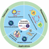
Fig. 1
Fig. 1. Schematic illustration of permeable polymersomes for diverse biomimetic applications.
Fig. 2
Fig. 2. (a) Schematic representation of a polystyrene-block-poly(l-isocyanoalanine (2-thiophen-3-ylethyl)amide) (PS-b-PIAT) polymersome. Yellow is PIAT and blue is PS. Reproduced with permission from ref. . Copyright 2003, Wiley-VCH. (b) The enzymatic activities of PS-b-PEG and PS-b-PIAT polymersome nanoreactors were assessed by the addition of H2O2. (I): PEG-b-PS, (II): PS-b-PIAT. Reproduced with permission from ref. . Copyright 2013, American Chemical Society.
Fig. 3
Fig. 3. (a) Schematic representation of poly(2-methyl-2-oxazoline)-block-poly(dimethylsiloxane)-block-(2-methyl-2-oxazoline) (PMOXA-b-PDMS-b-PMOXA) polymersomes as a functionalized nanoreactor. Reprinted from ref. . Copyright 2005, American Chemical Society. (b) Schematic representation of the nanoreactor system. (c) Channel- and pH-dependent nanoreactor activity in confocal microscopy (Magnification 400×; Zeiss ConfoCor 2, HAL 100, HBO 100, Software LSM 510 Zeiss): active nanoreactors with ompF pores show strong fluorescent activity at pH = 5 (I), but show no significant fluorescent activity without the ompF pores (II); active nanoreactors with ompF (III) and without ompF (IV) pores show no fluorescent activity at pH = 7.5. The substrate concentration in this experiment was 75 μM. Reprinted from ref. . Copyright 2006, American Chemical Society.
Fig. 4
Fig. 4. (a) Schematic representation of the formation of pH-responsive poly(ethylene oxide)-block-poly[2-(diethylamino) ethyl methacrylate-stat-3-(trimethoxysilyl) propyl methacrylate] [PEO-b-P(DEA-stat-TMSPMA)] polymersomes. (b) Variation of hydrodynamic vesicle diameter with solution pH: polymersomes prepared (I) with PEO43-b-P(DEA40-stat-TMSPMA40) and (II) without triethylamine; and (III) PEO43-b-P (DEA60-stat-TMSPMA)10, prepared with triethylamine. Reprinted from ref. . Copyright 2005, American Chemical Society. (c) Schematic representation of DOX/GOD@MCL-polymersomes as intelligent nanoreactors and drug delivery vehicles. Reprinted from ref. . Copyright 2020, Elsevier.
Fig. 5
Fig. 5. (a) DASA-functionalized visible light-responsive nanoreactors. Reprinted from ref. . Copyright 2018, American Chemical Society. (b) Amphiphilic poly(ethylene oxide)-block-poly(spiropyran) (PEO-b-PSPA) diblock copolymers self-assemble into polymersomes with hydrophobic bilayers containing carbamate-based hydrogen bonding motifs. Reprinted from ref. . Copyright 2015, American Chemical Society.
Fig. 6
Fig. 6. (a) Schematic representation of UV-responsive nanoreactors. (b) HRP activity test. (I–III) HRP-filled polymersomes before photoreaction with 2-hydroxy-4′-2-(hydroxyethoxy)-2-methylpropiophenone (PP-OH) and (IV–VII) after photoreaction with PP-OH. (IV) A-PMOXA-b-PDMS-b-PMOXA-A-HRP-PP-OH. (V) PMOXA-b-PDMS-b-PMOXA-HRP-PP-OH. (VI) PEO-b-PB-HRP-PP-OH. (VII) Free HRP. Reprinted from ref. . Copyright 2013, American Chemical Society. (c) The catalytic activity of the DASA esterase nanoreactor is activated by green light irradiation and deactivated by fading light irradiation. Reprinted from ref. . Copyright 2023, Springer Nature.
Fig. 7
Fig. 7. (a) Switchable chemical structural changes of poly(ethylene glycol)-block-poly(N-amidino) dodecyl acrylamide (PEG-b-PAD) copolymers. (b) Gas-controlled selective release of cargos from the polymersomes. Reprinted from ref. . Copyright 2013, Wiley-VCH.
Fig. 8
Fig. 8. (a) Schematic representation of conformation-regulated permeability of methoxy poly(ethylene glycol)-poly(l-cysteine) (mPEG-PLCC) polymersomes. (b) and hydrophilic drug R6G (c) from MPEG-PLCC assemblies in media with or without 10% H2O2. Reprinted from ref. . Copyright 2021, Wiley-VCH.
Fig. 9
Fig. 9. (a) Schematic representation of the emulsion-centrifugation process producing polymersomes. (b) The plot of vitro DOX release (weight%). Reprinted from ref. . Copyright 2013, American Chemical Society. (c) Schematic representation of the formation of permeable polymersomes utilizing pH–responsive and glucose–responsive block copolymers. Reprinted from ref. . Copyright 2009, Wiley-VCH.
Fig. 10
Fig. 10. (a) Reversible swelling–shrinking of hollow capsules upon switching between 25 and 40 °C at different pH. (b) Reversible swelling–shrinking of hollow capsules upon switching between pH = 6 and 8 at 25 °C. The diameter was measured by Dynamic Light Scattering (DLS). Reprinted from ref. . Copyright 2018, American Chemical Society.
Fig. 11
Fig. 11. (a) Preparation of the block copolymers. (b) Schematic representation of Meldrum's acid-based furan adduct (MELD) polymersomes and pyrazolone-based furan adduct (PYRA) polymersomes. (c) UV-vis absorbance indicates product formation upon light irradiation of HRP loaded into MELD polymersomes. (d) UV-vis absorbance indicating product formation upon light irradiation of GOx loaded into PYRA polymersomes. Continuous irradiation with white light (blue and pink), alternating light and dark cycle (orange), dark (black), and non-catalytic blank reaction (red). (e) Cascade pyrroliol reaction catalyzed by a mixture of MELD-HRP and PYRA-GOx nanoreactors, alternately deactivated in the dark and activated with red light (λ = 630 nm)(red), dark and green light (λ = 525 nm) (green), dark(black). Reprinted from ref. . Copyright 2018, American Chemical Society.
Fig. 12
Fig. 12. (a) Schematic representation of feedback-induced temporal control of polymersome nanoreactors. (b) Reversible nanoreactor “ON–OFF” regulation in time following repeated additions of urea. Reprinted from ref. . Copyright 2018, American Chemical Society.
Fig. 13
Fig. 13. (a) Schematic representation of polymersomes-in-polymersome microreactor. (b) Ensemble fluorescence measurements of the cascade reaction with all of the enzymes free in solution (▲), CalB in polystyrene-block-poly(l-isocyanoalanine (2-thiophen-3-ylethyl)amide) (PS-b-PIAT) nanoreactors (■), or CalB and ADH in PS-b-PIAT nanoreactors (O). (c) Ensemble fluorescence measurements of the cascade reaction with CalB in PS-b-PIAT nanoreactors (■), alcalase in PS-b-PIAT nanoreactors(Δ), free alcalase (●). Reprinted from ref. . Copyright 2014, Wiley-VCH. (d) The schematic representation of the enzymatic reaction of lipase substrate (DGGR) loaded on nano-particles and LipVes loaded with lipase enzyme, and these polymersomes are encapsulated as subcompartments into a polymer GUV. Reprinted from ref. . Copyright 2020, Wiley-VCH.
Fig. 14
Fig. 14. (a) Schematic representation of a hierarchical protocell. (b) Hierarchical protocell sequestering three kinds of proto-organelles loaded with different fluorescently labelled proteins. (c) Bulk fluorescence emission spectroscopy of self-assembled systems. BSA is labelled with fluorescent probes, which are quenched when BSA remains intact. Upon protease K (protK) activity fluorescence is observed. Encapsulation thus protects BSA from hydrolysis. Reprinted from ref. . Copyright 2019, American Chemical Society.
Fig. 15
Fig. 15. (a) Schematic representation of modified OmpF acting as a gate in catalytic nanocompartments. (b) Molecular representation of the OmpF-M cysteine mutant. (c) ZFE biodistribution in vivo and activity of AOs (lateral view of the ZFE crosssection). Blue: ZFE melanocytes. Green: HRP-Atto488. Red: RZLP. Reprinted from ref. . Copyright 2018, Nature Publishing Group.
Fig. 16
Fig. 16. (a) Schematic representation of the formation of molecular factories (MFs). (b) Left: Schematic representation of MFs containing AOs with OmpF. Right: Relative fluorescence intensity recordings of MFs containing AOs with OmpF. (c) Left: Schematic representation of MFs containing AOs without OmpF. Right: Relative fluorescence intensity recordings of MFs containing AOs without OmpF. (d) Schematic representation of ZFE injection and imaging. Reprinted from ref. . Copyright 2020, Wiley-VCH.
Fig. 17
Fig. 17. (a) Schematic representation of design and construction of the giant polymersomes (GPs). (b) UV-induced uptake of β-gal-FITC. (c) Fluorescence intensity of SP-GPs microreactors at different conditions. (d) Schematic representation of the coacervation process inside SP-GPs which could concentrate biomolecules. Reprinted from ref. . Copyright 2022, Wiley-VCH.
Fig. 18
Fig. 18. (a) Structure and composition of the amphiphilic diblock copolymer poly(oligo(ethylene glycol) monomethyl ether methacrylate-co-eosin Y)-block-poly(2-nitrobenzyloxycarbonylaminoethylmethacrylate) (P(OEGMA-co-EOS)-b-PNBOC) (POPN) and the hypoxia-activated prodrug/PEG ligand (AQ4N/PEG-UCNPs) polymersome assembly. (b) Optical transmission of the PEG-UCNPs polymersomes, depending on the time of NIR irradiation (in the presence of NIR (+)) and in the absence of (NIR (−)) irradiation. (c) Schematic representation of continuous release of the hydrophilic agent AQ4N in the polymersomes after NIR irradiation. Reprinted from ref. . Copyright 2021, American Chemical Society.
Fig. 19
Fig. 19. (a) Schematic representation of the emulsion-centrifugation process producing giant polymer vesosomes. The droplets containing poly(trimethylene carbonate)-block-poly(l-glutamic acid) (PTMC-b-PGA) which were poured onto the interface between toluene and 380 mOsm glucose aqueous solution passed through the interface by centrifugal forced and were packaged by PB-b-PEO polymersomes in toluene. (b) The plot of in vitro DOX release (weight%), (I) free DOX at 37 °C, (II) nano-DOX at 37 °C, (III) veso-DOX at 37 °C, (IV) nano-DOX at 20 °C and (V) veso-DOX at 20 °C. Reprinted from ref. . Copyright 2012, Wiley-VCH.
Fig. 20
Fig. 20. (a) Schematic representation of poly(N,N′-dimethylacrylamide)-block-poly(o-nitrobenzyl acrylate) (PDMA-b-PNBA) polymersomes for the release of drugs. (b) NR release spectra without UV irradiation. (c) NR emission spectra under UV irradiation. (d) DOX release upon UV irradiation at different times. Reprinted from ref. . Copyright 2020, Multidisciplinary Digital Publishing Institute.
Fig. 21
Fig. 21. (a) Schematic representation of SP-to-MC photochromic transition. (SP-to-MC: the transition from impermeable SP polymersomes to permeable MC polymersomes. SP: Spiropyran. MC: Zwitterionic merocyanine.) (b) The release of 5-dFu from SP and MC polymersomes upon UV irradiation. (c) Ladder-type controlled release profiles of 5-dFu from polymersomes under alternated UV-vis light irradiation. Reprinted from ref. . Copyright 2015, American Chemical Society.
Fig. 22
Fig. 22. (a) Schematic representation of TAT-polymersomes protecting the cell against H2O2. Scale bar = 100 nm. (b) Uptake of NBD-labelled polymersomes after 24 h at different polymersome concentrations (0.05–0.1 mg mL−1) and with different percentages of cell penetrating peptide (CPP) (0–5%). (c) The shielding effect of AONs on exogenous H2O2 in cells. CBOC and CBAC are both catalase. Reprinted from ref. . Copyright 2018, American Chemical Society.
Fig. 23
Fig. 23. (a) Schematic representation of PICsomes and enzyme@PICsomes via preformed PICsomes. Scale bar = 200 nm. (b) In vivo imaging of C26 tumor-bearing mice. The tumor site of the mouse was treated with (I) β-Gal@Cy5-PICsomes, (II) free β-Gal, and (III) HMDER-β-Gal. (c) The fluorescence imaging of organs ex vivo. Reprinted from ref. . Copyright 2016, Wiley-VCH.
Fig. 24
Fig. 24. (a) Schematic representation of glucose-responsive insulin delivery based on GOD-loaded methoxy poly(ethylene glycol)-poly(l-cysteine) (mPEG-PLCC) polymersomes. (b) Blood glucose levels in STZ-induced diabetic mice after PBS, E + I@MPEG-PLCC, I@MPEG-PLCC, and insulin injection at different times. (c) Plasma human insulin levels in STZ-induced diabetic mice after E + I@MPEG-PLCC and insulin injection for different times. Reprinted from ref. . Copyright 2021, Wiley-VCH.
Fig. 25
Fig. 25. A concluding scheme for the challenges of permeable polymersomes.

Similar articles

Cited by

References

    1. Fong C. Le T. Drummond C. J. Chem. Soc. Rev. 2012;41:1297–1322. doi: 10.1039/C1CS15148G. - DOI - PubMed
    1. Li H. Carter J. D. LaBean T. H. Mater. Today. 2009;12:24–32. doi: 10.1016/S1369-7021(09)70157-9. - DOI
    1. Klok H. A. Lecommandoux S. Adv. Mater. 2001;13:1217–1229. doi: 10.1002/1521-4095(200108)13:16<1217::AID-ADMA1217>3.0.CO;2-D. - DOI
    1. Elsabahy M. Heo G. S. Lim S. M. Sun G. R. Wooley K. L. Chem. Rev. 2015;115:10967–11011. doi: 10.1021/acs.chemrev.5b00135. - DOI - PMC - PubMed
    1. Tu Y. Peng F. Adawy A. Men Y. Abdelmohsen L. K. E. A. Wilson D. A. Chem. Rev. 2016;116:2023–2078. doi: 10.1021/acs.chemrev.5b00344. - DOI - PubMed