. 2023 Jun 16;14(27):7524-7536.
doi: 10.1039/d3sc00728f. eCollection 2023 Jul 12.

Modulation of IL-17 backbone dynamics reduces receptor affinity and reveals a new inhibitory mechanism

Affiliations

Modulation of IL-17 backbone dynamics reduces receptor affinity and reveals a new inhibitory mechanism

Daniel J Shaw et al. Chem Sci. .

Abstract

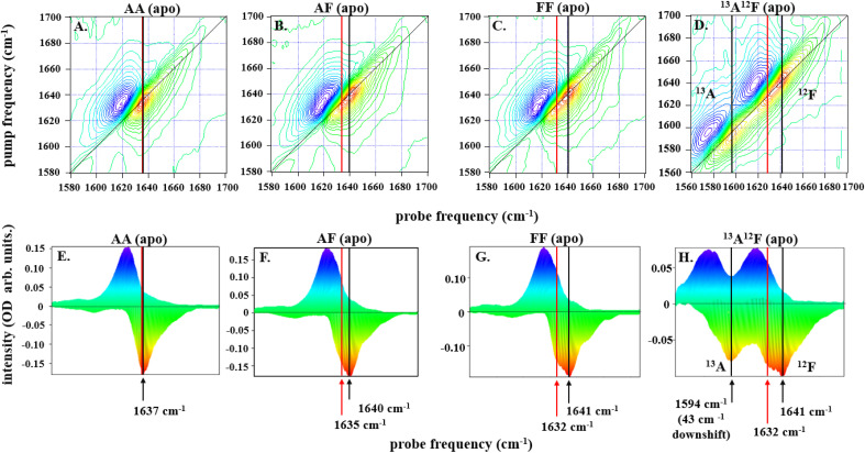
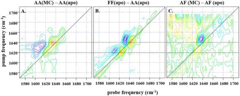
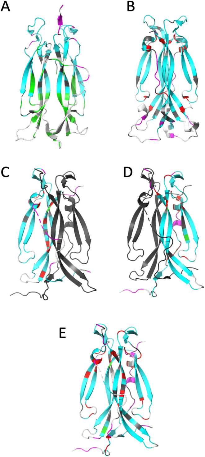
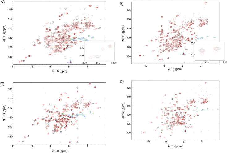
Knowledge of protein dynamics is fundamental to the understanding of biological processes, with NMR and 2D-IR spectroscopy being two of the principal methods for studying protein dynamics. Here, we combine these two methods to gain a new understanding of the complex mechanism of a cytokine:receptor interaction. The dynamic nature of many cytokines is now being recognised as a key property in the signalling mechanism. Interleukin-17s (IL-17) are proinflammatory cytokines which, if unregulated, are associated with serious autoimmune diseases such as psoriasis, and although there are several therapeutics on the market for these conditions, small molecule therapeutics remain elusive. Previous studies, exploiting crystallographic methods alone, have been unable to explain the dramatic differences in affinity observed between IL-17 dimers and their receptors, suggesting there are factors that cannot be fully explained by the analysis of static structures alone. Here, we show that the IL-17 family of cytokines have varying degrees of flexibility which directly correlates to their receptor affinities. Small molecule inhibitors of the cytokine:receptor interaction are usually thought to function by either causing steric clashes or structural changes. However, our results, supported by other biophysical methods, provide evidence for an alternate mechanism of inhibition, in which the small molecule rigidifies the protein, causing a reduction in receptor affinity. The results presented here indicate an induced fit model of cytokine:receptor binding, with the more flexible cytokines having a higher affinity. Our approach could be applied to other systems where the inhibition of a protein-protein interaction has proved intractable, for example due to the flat, featureless nature of the interface. Targeting allosteric sites which modulate protein dynamics, opens up new avenues for novel therapeutic development.


Conflict of interest statement

The authors declare the following financial interests/personal relationships which may be considered as potential competing interests: Daniel J. Shaw, Monika-Sarah E. D. Schulze, Christine E. Prosser, Alastair D. G. Lawson, Alistair J. Henry and Richard J. Taylor are or have been employees of UCB and may hold UCB shares and/or stock options.

Figures

Fig. 1
Fig. 1. Ribbon representation of IL-17AA illustrating major structural elements (generated using PDB code: 7UWM). The illustration shows one protomer to the left and the other to the right of the dotted line resulting, in the case of the homo-dimers, a symmetric dimer.
Fig. 2
Fig. 2. 2D-IR spectra for all dimers, IL-17AA in panels A and E, IL-17AF in panels B and F, IL-17FF in panels C and G, and IL-17AF13A12F in panels D and H, as labelled at a pump–probe delay time (Tw) of 250 fs are depicted along the top row of (A–D). The color scale runs from red (negative) to blue (positive). Diagonal projections of the spectra in A–D are shown in E–H. The frequency positions of the main band and shoulder described in the text are marked with arrows (black: main band; red, shoulder). The 13A12F sample contains IL17AF in which the A protomer has been isotopically enriched with 13C to shift the amide I band to lower frequency and remove overlap between the amide I bands of the A and F protomers.
Fig. 3
Fig. 3. (A) shows the difference spectrum obtained by subtraction of IL-17AA apo-2D-IR data from a 1 : 2 macrocycle (MC) bound sample at waiting time (Tw) of 250 fs and at parallel polarisation, whilst (B) shows the subsequent difference obtained from subtraction of the same IL-17AA apo-data from that for an IL17-FF apo-sample at the same waiting time. A similar difference is shown in (C), obtained for an AF macrocycle minus AF apo-subtraction, again at a Tw of 250 fs, but with a much lower signal-to-noise level that is indicative of the low binding affinity of the AF isoform. The dashed horizontal lines highlight the similarity in peak positions for both subtractions, indicating that the binding of the macrocycle to both IL-17AA and IL-17AF induces changes that are much more comparable both spectroscopically and structurally to the IL-17FF apo-form.
Fig. 4
Fig. 4. Ribbon representation of IL-17 isoforms. (A) IL-17AA (PDB code 4HSA), (B) IL-17FF (PDB code 1JPY), (C) IL-17Af (PDB code 5N92), (D) IL-17Fa and (E) IL-17AF:MC complex. The backbone is colour coded by motion type. Colours are, white – not assigned, grey – overlapped resonance therefore no reliable data, cyan – relaxation dominated by overall correlation time (τc), magenta – motions faster (τe) than the overall correlation time (τc) dominate relaxation, green – resonance decayed too quickly to determine R2 and therefore are interpreted as undergoing conformational exchange (Rex), red – exchange contribution (Rex) to relaxation of resonance. Due to the absence of a significant portion of the long interface coil region in the crystal structure of free IL-17AA, the more complete structure obtained in complex with IL-17RA was used.
Fig. 5
Fig. 5. 15N-trosy spectra of the IL-17 isoforms in the presence and absence of MC. Black contours are apo protein and red contours are in the presence of MC, green/blue contours are aliased arginine sidechain resonances. (A) IL-17AA (B) IL-17FF (C) 15N labelled A protomer of IL-17AF (D) 15N labelled F protomer of IL-17AF. Insert panel A shows additional signals due to de-symmetrisation of the Il-17AA dimer. and the insert to panel B shows additional signals resulting from incomplete saturation of the IL-17FF homodimer.

Similar articles

Cited by

References

    1. Krueger J. G. Wharton, Jr K. A. Schlitt T. Suprun M. Torene R. I. Jiang X. Wang C. Q. Fuentes-Duculan J. Hartmann N. Peters T. Koroleva I. Hillenbrand R. Letzkus M. Yu X. Li Y. Glueck A. Hasselberg A. Flannery B. Suarez-Farinas M. Hueber W. J. Allergy Clin. Immunol. 2019;144:750–763. doi: 10.1016/j.jaci.2019.04.029. - DOI - PubMed
    1. Reich K. Papp K. A. Matheson R. T. Tu J. H. Bissonnette R. Bourcier M. Gratton D. Kunynetz R. A. Poulin Y. Rosoph L. A. Stingl G. Bauer W. M. Salter J. M. Falk T. M. Blodorn-Schlicht N. A. Hueber W. Sommer U. Schumacher M. M. Peters T. Kriehuber E. Lee D. M. Wieczorek G. A. Kolbinger F. Bleul C. C. Exp. Dermatol. 2015;24:529–535. doi: 10.1111/exd.12710. - DOI - PMC - PubMed
    1. Krueger J. G. Fretzin S. Suarez-Farinas M. Haslett P. A. Phipps K. M. Cameron G. S. McColm J. Katcherian A. Cueto I. White T. Banerjee S. Hoffman R. W. J. Allergy Clin. Immunol. 2012;130:145–154. doi: 10.1016/j.jaci.2012.04.024. - DOI - PMC - PubMed
    1. Liu L. Lu J. Allan B. W. Tang Y. Tetreault J. Chow C. K. Barmettler B. Nelson J. Bina H. Huang L. Wroblewski V. J. Kikly K. J. Inflammation Res. 2016;9:39–50. doi: 10.2147/JIR.S100940. - DOI - PMC - PubMed
    1. Reich K. Warren R. B. Lebwohl M. Gooderham M. Strober B. Langley R. G. Paul C. De Cuyper D. Vanvoorden V. Madden C. Cioffi C. Peterson L. Blauvelt A. N. Engl. J. Med. 2021;385:142–152. doi: 10.1056/NEJMoa2102383. - DOI - PubMed