

Whole mandibular canal segmentation using transformed dental CBCT volume in Frenet frame
- PMID: 37449128
- PMCID: PMC10336514
- DOI: 10.1016/j.heliyon.2023.e17651
Whole mandibular canal segmentation using transformed dental CBCT volume in Frenet frame
Abstract
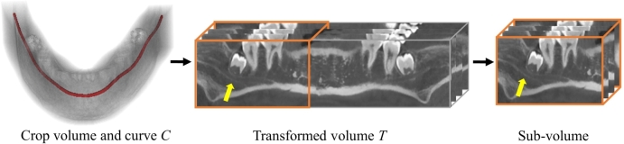
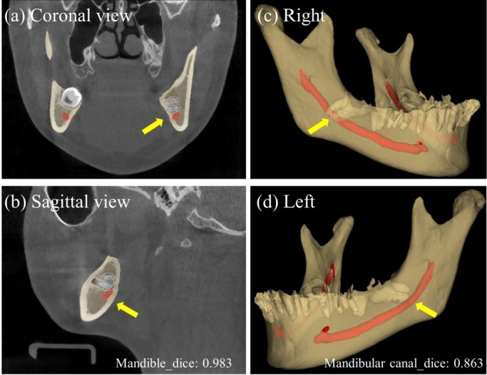
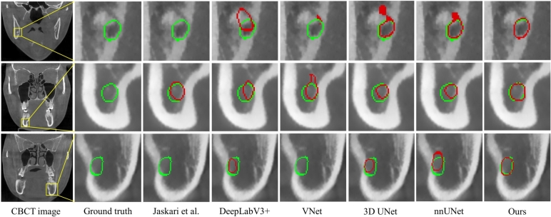
Accurate segmentation of the mandibular canal is essential in dental implant and maxillofacial surgery, which can help prevent nerve or vascular damage inside the mandibular canal. Achieving this is challenging because of the low contrast in CBCT scans and the small scales of mandibular canal areas. Several innovative methods have been proposed for mandibular canal segmentation with positive performance. However, most of these methods segment the mandibular canal based on sliding patches, which may adversely affect the morphological integrity of the tubular structure. In this study, we propose whole mandibular canal segmentation using transformed dental CBCT volume in the Frenet frame. Considering the connectivity of the mandibular canal, we propose to transform the CBCT volume to obtain a sub-volume containing the whole mandibular canal based on the Frenet frame to ensure complete 3D structural information. Moreover, to further improve the performance of mandibular canal segmentation, we use clDice to guarantee the integrity of the mandibular canal structure and segment the mandibular canal. Experimental results on our CBCT dataset show that integrating the proposed transformed volume in the Frenet frame into other state-of-the-art methods achieves a improvement in Dice performance. Our proposed method can achieve impressive results with a Dice value of 0.865 (±0.035), and a clDice value of 0.971 (±0.020), suggesting that our method can segment the mandibular canal with superior performance.
Keywords: CBCT; Deep learning; Frenet frame; Mandibular canal; Segmentation.
© 2023 The Author(s).
Conflict of interest statement
The authors declare that there is no conflict of interest regarding the publication of this paper.
Figures


References
-
- Yu S.-K., Lee M.-H., Jeon Y.H., Chung Y.Y., Kim H.-J. Anatomical configuration of the inferior alveolar neurovascular bundle: a histomorphometric analysis. Surg. Radiol. Anat. 2016;38(2):195–201. - PubMed
-
- Ebenezer V., Sumathi G., et al. Importance of cone beam computed tomography in dental implants: a review. J. Positive Sch. Psychol. 2022;6(3):3798–3800.
-
- de Oliveira-Santos C., Souza P.H.C., de Azambuja Berti-Couto S., Stinkens L., Moyaert K., Rubira-Bullen I.R.F., Jacobs R. Assessment of variations of the mandibular canal through cone beam computed tomography. Clin. Oral Investig. 2012;16(2):387–393. - PubMed
-
- Iwanaga J., Katafuchi M., Matsushita Y., Kato T., Horner K., Tubbs R.S. Anatomy of the mandibular canal and surrounding structures: part I: morphology of the superior wall of the mandibular canal. Ann. Anat. 2020;232 - PubMed
-
- Iwanaga J., Shiromoto K., Kato T., Tanaka T., Ibaragi S., Tubbs R.S. Anatomy of the mandibular canal and surrounding structures. Part II: cancellous pattern of the mandible. Ann. Anat. 2020;232 - PubMed
LinkOut - more resources
Full Text Sources
