Review
. 2023 Aug 11;67(4):685-699.
doi: 10.1042/EBC20220257.

Mechanisms of bioleaching: iron and sulfur oxidation by acidophilic microorganisms

Affiliations
Review

Mechanisms of bioleaching: iron and sulfur oxidation by acidophilic microorganisms

Sarah Jones et al. Essays Biochem. .

Abstract

Bioleaching offers a low-input method of extracting valuable metals from sulfide minerals, which works by exploiting the sulfur and iron metabolisms of microorganisms to break down the ore. Bioleaching microbes generate energy by oxidising iron and/or sulfur, consequently generating oxidants that attack sulfide mineral surfaces, releasing target metals. As sulfuric acid is generated during the process, bioleaching organisms are typically acidophiles, and indeed the technique is based on natural processes that occur at acid mine drainage sites. While the overall concept of bioleaching appears straightforward, a series of enzymes is required to mediate the complex sulfur oxidation process. This review explores the mechanisms underlying bioleaching, summarising current knowledge on the enzymes driving microbial sulfur and iron oxidation in acidophiles. Up-to-date models are provided of the two mineral-defined pathways of sulfide mineral bioleaching: the thiosulfate and the polysulfide pathway.

Keywords: Acidophiles; Bioleaching; Iron oxidation; Sulfur oxidation.


Conflict of interest statement

The authors declare that there are no competing interests associated with the manuscript.

Figures

Figure 1
Figure 1. Simplified overview of the bioleaching process showing the regeneration of oxidants by iron- and sulfur- oxidising microbes, resulting in the release of target metals
Figure 2
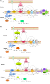
Figure 2. Model of sulfur oxidation in the Acidithiobacilli
Model of sulfur oxidation in (A) A. ferrooxidans; (B) A. thiooxidans and (C) A. ferrivorans. Sulfide oxidation proceeds via the inner membrane-bound sulfide-quinone reductase (SQR), which facilitates the oxidation of hydrogen sulfide to elemental sulfur. Insoluble elemental sulfur in the periplasm is most likely converted to glutathionate persulfide (GSSH) by membrane bound thiols prior to oxidation. This GSSH is transported via transferases (DsrE, TusA and Rhd) to a heterodisulfide reductase (HDR) complex, which catalyses its oxidation to sulfite and GSH. Alternatively, elemental sulfur may be oxidised by sulfur oxygenase reductase (SOR) or sulfur dioxygenase (SDO). It is predicted that sulfite oxidation in A. ferrooxidans and A. ferrivorans is catalysed by an as-yet unknown enzyme, generating adenosine-5-phosphosulfate (APS), which is then further oxidised to sulfate, with concomitant ATP and proton generation by sulfate adenylyltransferase (SAT). In A. thiooxidans, sulfite oxidation occurs via phosphoadenosine phosphosulfate (PAPS) reductase, where sulfite is first oxidised to PAPS by the PAPS reductase, then oxidised to APS, and sulfate by APS kinase. In all three species, the oxidation of thiosulfate to tetrathionate is mediated by thiosulfate quinone oxidoreductase (TQO) or thiosulfate dehydrogenase (TSD), while an outer membrane-bound, homodimeric tetrathionate hydrolase (TetH) hydrolyses tetrathionate to thiosulfate. A. thiooxidans and A. ferrivorans both possess the alternative sulfur oxidation pathway, SOX. Across the Acidithiobacilli, electrons produced by RISC oxidation are thought to be transferred to the quinone pool (Q/QH2), from which they are transported to the membrane bound terminal oxidases bo3 and/or bd. Alternately, the electrons generated in RISC oxidation can be transferred indirectly to an aa3 oxidase for O2 reduction (via high potential iron-sulfur protein (HiPIP)), or to a NADH1 complex, via which NADH can be generated. These figures were created based on information collected [16–30].
Figure 3
Figure 3. Schematic of Sox clusters present in Acidithiobacillus species
(A) Sox I cluster present in At. thiooxidans. Adapted from [27] and (B) Sox II cluster present in At. thiooxidans and At. ferrivorans. Adapted from [21].
Figure 4
Figure 4. A. ferrooxidans ferrous iron oxidation electron transfer model
The electron transport chain in A. ferroxidans spans the inner (IM) and outer membranes (OM), forming a super-complex that begins with a high molecular-weight outer membrane bound cytochrome c (Cyc2). Iron remains outside the cell as it is oxidised via CycC. Electrons flow from CycC to the periplasmic protein rusticyanin (Rus) and are thereafter directed to either the downhill pathway or the uphill pathway. In the downhill pathway, electrons move from Rus to the membrane-bound periplasmic cytochrome c, Cyc1, finally reducing oxygen via aa3-type terminal cytochrome oxidase. In the uphill pathway, electrons move from Rus to the alternate membrane-bound periplasmic cytochrome c, CycA1. From CycA1, electrons pass to a reverse-functioning bc1 complex positioned within the inner cell membrane and then via the membrane-associated ubiquinone pool to the NADH oxidoreductase complex (NDH1), where NAD+ is reduced. The hypothetical gene cup (previously ORF1), appears in the rus gene operon. The role of Cup is as yet undetermined, but has been speculated to include delivering copper to aa3and/or Rus, or facilitating electron transfer between Cyc2 and Cyc1 excluding Rus (Figure based on information and diagrams in: [17,18,34,38,39,41,44])
Figure 5
Figure 5. Schematics of operons associated with iron oxidation in the Acicidithiobacilli
(A) The rus operon. Promoters are indicated by black arrows. Two promoters are present upstream of cyc2 which encodes the outer membrane-bound CycC. The aa3 subunit is encoded by coxABCD. Based on images and information in [23,35]. [23] (B) The cta operon. White indicates that the gene is not represented on electron transport models. Figure based on information in [19] (C) The petI operon. Promoter is shown by the black arrow [48,50].
Figure 6
Figure 6. Model of iron oxidation in L. ferrodiazotrophum
Direct oxidation occurs via the outer membrane Cyt572, with electrons passing through a potential periplasmic Cyt579 to cytochrome c to inner membrane bound terminal oxidases. Based on images and information in [37,58].
Figure 7
Figure 7. Model of iron oxidation in F. acidarmanus
Oxidation of iron is via a sulfocyanin-type blue-copper protein, the exact location of which remains speculative. SDH: succinate dehydrogenase. Based on images and information in [37,59,60].
Figure 8
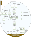
Figure 8. Model of chalcopyrite dissolution via the polysulfide pathway
PSR: polysulfide reductase, SQR: sulfide-quinone reductase, SOR: sulfur oxygenase reductase, SDO: sulfur dioxygenase, HDR: heterodisulfide reductase, SAT: sulfate adenylyltransferase, TetH: tetrathionate hydrolase, TQO: thiosulfate-quinone oxidoreductase, TSD: thiosulfate dehydrogenase, SOX: sulfur oxidation pathway, RUS: rusticyanin, SoxE: Sulfocyanin, IRO: high potential iron-sulfur protein, Cyt579: Cytochrome 572.
Figure 9
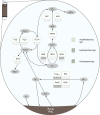
Figure 9. Model of pyrite dissolution via the thiosulfate pathway
SQR: sulfide-quinone reductase, SOR: sulfur oxygenase reductase, SDO: sulfur dioxygenase, HDR: heterodisulfide reductase, SAT: sulfate adenylyltransferase, TetH: tetrathionate hydrolase, TQO: thiosulfate-quinone oxidoreductase, TSD: thiosulfate dehydrogenase, SOX: sulfur oxidation pathway, RUS: rusticyanin, SoxE: Sulfocyanin, IRO: high potential iron-sulfur protein, Cyt579: Cytochrome 572.

Similar articles

Cited by

References

    1. Zhao H., Zhang Y., Zhang X., Qian L., Sun M., Yang Y.et al. (2019) The dissolution and passivation mechanism of chalcopyrite in bioleaching: An overview. Minerals Engineering, vol. 136, pp. 140–154, Elsevier Ltd; 10.1016/j.mineng.2019.03.014 - DOI
    1. Bini E. (2010) Archaeal transformation of metals in the environment. FEMS Microbiol. Ecol. 73, 1–16 10.1111/j.1574-6941.2010.00876.x - DOI - PubMed
    1. Valdés J., Cárdenas J.P., Quatrini R., Esparza M., Osorio H., Duarte F.et al. (2010) Comparative genomics begins to unravel the ecophysiology of bioleaching. Hydrometallurgy 104, 471–476 10.1016/j.hydromet.2010.03.028 - DOI
    1. Shiers D.W., Collinson D.M. and Watling H.R. (2016) Life in heaps: a review of microbial responses to variable acidity in sulfide mineral bioleaching heaps for metal extraction. Res. Microbiol. 167, 576–586 10.1016/j.resmic.2016.05.007 - DOI - PubMed
    1. Rohwerder T., Gehrke T., Kinzler K. and Sand W. (2003) Bioleaching review part A: Progress in bioleaching: Fundamentals and mechanisms of bacterial metal sulfide oxidation. Appl. Microbiol. Biotechnol. 63, 239–248 10.1007/s00253-003-1448-7 - DOI - PubMed

Publication types