. 2023 Dec;18(12):2711-2719.
doi: 10.4103/1673-5374.374654.

Quantitative proteomic and phosphoproteomic analyses of the hippocampus reveal the involvement of NMDAR1 signaling in repetitive mild traumatic brain injury

Affiliations

Quantitative proteomic and phosphoproteomic analyses of the hippocampus reveal the involvement of NMDAR1 signaling in repetitive mild traumatic brain injury

Zhicheng Tian et al. Neural Regen Res. 2023 Dec.

Abstract

The cumulative damage caused by repetitive mild traumatic brain injury can cause long-term neurodegeneration leading to cognitive impairment. This cognitive impairment is thought to result specifically from damage to the hippocampus. In this study, we detected cognitive impairment in mice 6 weeks after repetitive mild traumatic brain injury using the novel object recognition test and the Morris water maze test. Immunofluorescence staining showed that p-tau expression was increased in the hippocampus after repetitive mild traumatic brain injury. Golgi staining showed a significant decrease in the total density of neuronal dendritic spines in the hippocampus, as well as in the density of mature dendritic spines. To investigate the specific molecular mechanisms underlying cognitive impairment due to hippocampal damage, we performed proteomic and phosphoproteomic analyses of the hippocampus with and without repetitive mild traumatic brain injury. The differentially expressed proteins were mainly enriched in inflammation, immunity, and coagulation, suggesting that non-neuronal cells are involved in the pathological changes that occur in the hippocampus in the chronic stage after repetitive mild traumatic brain injury. In contrast, differentially expressed phosphorylated proteins were mainly enriched in pathways related to neuronal function and structure, which is more consistent with neurodegeneration. We identified N-methyl-D-aspartate receptor 1 as a hub molecule involved in the response to repetitive mild traumatic brain injury , and western blotting showed that, while N-methyl-D-aspartate receptor 1 expression was not altered in the hippocampus after repetitive mild traumatic brain injury, its phosphorylation level was significantly increased, which is consistent with the omics results. Administration of GRP78608, an N-methyl-D-aspartate receptor 1 antagonist, to the hippocampus markedly improved repetitive mild traumatic brain injury-induced cognitive impairment. In conclusion, our findings suggest that N-methyl-D-aspartate receptor 1 signaling in the hippocampus is involved in cognitive impairment in the chronic stage after repetitive mild traumatic brain injury and may be a potential target for intervention and treatment.

Keywords: Grin1; N-methyl-D-aspartate; N-methyl-D-aspartate receptor 1; cognitive impairment; hippocampus; learning; memory; phosphoproteomic; proteomic; repetitive mild traumatic brain injury (rmTBI); secondary injury.


Conflict of interest statement

None

Figures
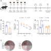
Figure 1
Figure 1
rmTBI induced delayed neurological dysfunction in the hippocampus. (A) Experimental design. (B) Novel object recognition index in the sham group (n = 6) and in the rmTBI group after injury (n = 6) (unpaired t-test, t = 2.306, P = 0.0438). (C–E) Morris water maze test (n = 6). Latency to find the platform (C) during the 4-day training phase (two-way analysis of variance, F = 23.80, P = 0.0006), average time spent in quadrants (Q1–3: the 1st–3rd quadrant; Q4: the 4th quadrant) (D) (two-way analysis of variance, F = 6.791, shamQ1–3 vs. shamQ4: P = 0.0014, rmTBIQ1–3 vs. rmTBIQ4: P = 0.9859, shamQ4 vs. rmTBIQ4: P = 0.0115) and representative traces (E) of swimming trajectories in the sham group and rmTBI group in the test phase. Q4 is the target quadrant. All data are expressed as the mean ± SEM. *P < 0.05, **P < 0.01, ***P < 0.001. MWM: Morris water maze; NOR: novel object recognition; ns: not significant; rmTBI: repetitive mild traumatic brain injury.
Figure 2
Figure 2
rmTBI induces structural lesions in the hippocampus. (A) Representative images of terminal deoxynucleotidyl transferase dUTP nick-end labeling (TUNEL) staining and relative integrated optical density (IOD) of TUNEL signals (n = 3) (unpaired t test, t = 0.2743, P = 0.7974). Blue: 4′,6-diamidino-2-phenylindole (DAPI), nucleus; green: TUNEL, apoptotic cells. (B and C) Immunostaining with an anti-p-tau antibody. Representative images with p-tau-positive staining (B) and the relative IOD of p-tau staining (C) in the hippocampus in the sham and rmTBI groups (six sections from three mice (two sections per mouse) from each group) (unpaired t-test, t = 6.218, P < 0.0001). Blue: DAPI, nucleus; green: p-tau. (D) Representative image and software-remodeled drawing of Golgi staining. (E) Total density of dendritic spines in hippocampal neurons in the sham group and in the rmTBI group after injury (unpaired t-test, t = 5.762, P < 0.0001), as well as the average volume of spines in two groups (unpaired t-test, t = 0.818, P = 0.4299). (F) Stubby spine density (unpaired t-test, t = 2.911, P = 0.0073) and mushroom spine density (Mann-Whitney U test, P = 0.0287) in hippocampal neurons in the sham group and in the rmTBI group after injury. Data are expressed as the mean ± SEM. *P < 0.05, **P < 0.01, ****P < 0.0001. ns: Not significant; rmTBI: repetitive mild traumatic brain injury.
Figure 3
Figure 3
Proteomic analysis of hippocampal tissue in a mouse model of rmTBI. (A) Number of differentially expressed proteins (DEPs) in the hippocampus. (B) Volcano plot of DEPs in the hippocampus. (C) Heatmap of selected DEPs. Light red represents high expression, while light blue represents low expression. (D and E) Bubble chart of GO enrichment analysis of upregulated (D) and downregulated (E) proteins. The bubbles display the terms in each GO category, including BP, CC, and MF. (F and G) Bubble chart of KEGG pathway analysis results for upregulated (F) and downregulated (G) proteins. The bubbles display the KEGG pathway terms. (H) Bubble chart of domains identified in the upregulated proteins. (I) DEP protein-protein interaction network. Red represents upregulated proteins, while green represents downregulated proteins. BP: Biological process; CC: cellular component; GO: Gene Ontology; HPC: hippocampus; KEGG: Kyoto Encyclopedia of Genes and Genomes; MF: molecular function; rmTBI: repetitive mild traumatic brain injury.
Figure 4
Figure 4
Phosphoproteomic analysis of hippocampal tissue in a mouse model of rmTBI. (A) Number of differentially expressed phosphorylated sites (DEPSs) and differentially expressed phosphorylated proteins (DEPPs). (B) Volcano plot of DEPSs in the hippocampus. (C) Heatmap of partial DEPSs. Light red represents high expression, while light blue represents low expression. (D and E) Bubble chart of GO enrichment analysis results for upregulated (D) and downregulated (E) phosphorylated proteins. The GO categories include BP, CC, and MF. (F and G) Bubble chart of KEGG pathway analysis results for upregulated (F) and downregulated (G) phosphorylated proteins. (H and I) Bubble chart of the domains identified in upregulated (H) and downregulated (I) phosphorylated proteins. The bubbles display terms corresponding to each GO category, KEGG pathway, or domain. (J) DEPP PPI network and clusters. The bar blot displays the terms associated with each cluster. BP: Biological process; CC: cellular component; GO: Gene Ontology; KEGG: Kyoto Encyclopedia of Genes and Genomes; MF: molecular function; PPI: protein-protein interaction; rmTBI: repetitive mild traumatic brain injury.
Figure 5
Figure 5
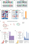
Analysis of phosphorylation motifs and upstream kinases. (A) Serine motifs with the maximal (11) and minimal (49) fold increase. (B) Threonine motifs with the maximal (1) and minimal (12) fold increase. (C and D) Heatmaps of phosphorylated sites around serine (C) and threonine (D) residues. Red represents high frequency, and blue represents low frequency. Each letter represents an amino acid. (E) Number of predicted upstream kinases. (F) The interaction network of upstream kinases and substrates. (G and H) UpSet plots of GO (G) and KEGG (H) pathways. The bar plots and table are arranged from highest to lowest degree of intersection. The tables display the name of the intersection gene. GO: Gene Ontology; HPC: hippocampus; KEGG: Kyoto Encyclopedia of Genes and Genomes; rmTBI: repetitive mild traumatic brain injury.
Figure 6
Figure 6
NMDAR1 helps regulate hippocampus-mediated cognitive dysfunction after rmTBI. (A) Western blot of hippocampal NMDAR1 and pNMDAR1. No change in NMDAR1 protein expression was detected (unpaired t-test, t = 1.406, P = 0.2324), and a relative increase in pNMDAR1 protein expression (unpaired t-test, t = 2.827, P = 0.0475) was detected (n = 3). NMDAR1: 105 kDa, pNMDAR1: 105 kDa, Tubulin: 55 kDa. (B) Schematic showing how saline and CGP78608 were injected into the hippocampus. (C) The decreased NOR index induced by rmTBI was almost abolished by treatment with CGP78608, but not saline (n = 6) (unpaired t-test, t = 3.168, P = 0.0100). (D) Interaction network among Grin1 and its upstream kinases. (E) Grin1 PPI network. All data are expressed as the mean ± SEM. *P < 0.05. NMDAR1: N-methyl-D-aspartate receptor 1; NOR: novel object recognition; pNMDAR1: phosphorylation N-methyl-D-aspartate receptor 1; rmTBI: repetitive mild traumatic brain injury.

Similar articles

Cited by

References

    1. Anacker C, Hen R. Adult hippocampal neurogenesis and cognitive flexibility - linking memory and mood. Nat Rev Neurosci. 2017;18:335–346. - PMC - PubMed
    1. Apweiler R, Biswas M, Fleischmann W, Kanapin A, Karavidopoulou Y, Kersey P, Kriventseva EV, Mittard V, Mulder N, Phan I, Zdobnov E. Proteome analysis database:online application of interpro and clustr for the functional classification of proteins in whole genomes. Nucleic Acids Res. 2001;29:44–48. - PMC - PubMed
    1. Aungst SL, Kabadi SV, Thompson SM, Stoica BA, Faden AI. Repeated mild traumatic brain injury causes chronic neuroinflammation, changes in hippocampal synaptic plasticity, and associated cognitive deficits. J Cereb Blood Flow Metab. 2014;34:1223–1232. - PMC - PubMed
    1. Ayubcha C, Revheim ME, Newberg A, Moghbel M, Rojulpote C, Werner TJ, Alavi A. A critical review of radiotracers in the positron emission tomography imaging of traumatic brain injury:fdg, tau, and amyloid imaging in mild traumatic brain injury and chronic traumatic encephalopathy. Eur J Nucl Med Mol Imaging. 2021;48:623–641. - PubMed
    1. Bader M, Li Y, Tweedie D, Shlobin NA, Bernstein A, Rubovitch V, Tovar-Y-Romo LB, Dimarchi RD, Hoffer BJ, Greig NH, Pick CG. Neuroprotective effects and treatment potential of incretin mimetics in a murine model of mild traumatic brain injury. Front Cell Dev Biol. 2019;7:356. - PMC - PubMed