Review
. 2023 Jul 14;72(3):301-307.
doi: 10.33549/physiolres.935007.

"Double-edged sword" effect of reactive oxygen species (ROS) in tumor development and carcinogenesis

Affiliations
Review

"Double-edged sword" effect of reactive oxygen species (ROS) in tumor development and carcinogenesis

W Zhao et al. Physiol Res. .

Abstract

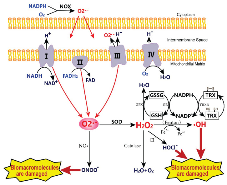
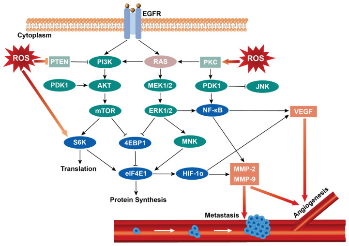
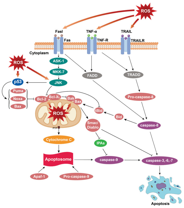
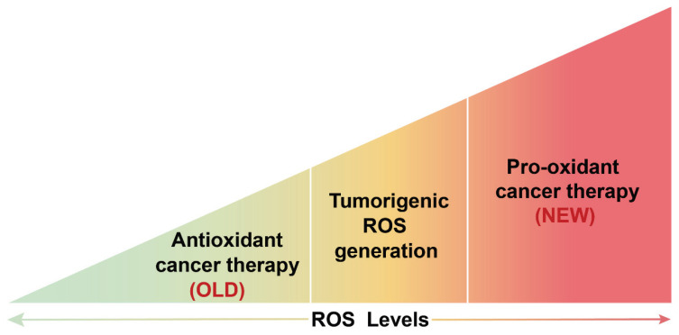
Reactive oxygen species (ROS) are small reactive molecules produced by cellular metabolism and regulate various physiological and pathological functions. Many studies have shown that ROS plays an essential role in the proliferation and inhibition of tumor cells. Different concentrations of ROS can have a "double-edged sword" effect on the occurrence and development of tumors. A certain concentration of ROS can activate growth-promoting signals, enhance the proliferation and invasion of tumor cells, and cause damage to biomacromolecules such as proteins and nucleic acids. However, ROS can enhance the body's antitumor signal at higher levels by initiating oxidative stress-induced apoptosis and autophagy in tumor cells. This review analyzes ROS's unique bidirectional regulation mechanism on tumor cells, focusing on the key signaling pathways and regulatory factors that ROS affect the occurrence and development of tumors and providing ideas for an in-depth understanding of the mechanism of ROS action and its clinical application.


Conflict of interest statement

Conflict of Interest

There is no conflict of interest.

Figures

Fig. 1
Fig. 1
The formation and metabolic mechanism of ROS. The O2•− produced by NADPH oxidase and mitochondrial electron transport chain can react with NO• or be catalyzed by SOD. H2O2 can be converted into H2O by antioxidant detoxification substances in mitochondria or cytoplasm and can also be generated into OH by the Fenton reaction. Toxic metabolites such as ONOO and OH cause damage to biological macromolecules, apoptosis, and autophagy. The GSH, as a part of Glutathione peroxidase and TRX systems, are antioxidant enzymes that catalyze the efficient decomposition of H2O2. I-IV: mitochondrial complexes I to IV; NOX: NADPH oxidase; SOD: superoxide dismutase; GSSG: glutathione disulfide; GSH: glutathione; TRX: thioredoxin; TRXR: thioredoxin reductase.
Fig. 2
Fig. 2
The relationship between ROS and angiogenesis and tumor metastasis. The increase of endogenous or exogenous ROS levels can affect the progress of tumor cells in many ways, such as releasing downstream growth factors and cytokines through PI3K/Akt/mTOR, RAS/Raf/MAPK, and other signal pathways, promoting the up-regulation of HIF-1α, VEGF and MMPs expression, activating NF-κB signal to cause angiogenesis and starting EMT to induce tumor invasion and migration. EGFR: epidermal growth factor receptor; PTEN: phosphatase and tensin homolog deleted on chromosome ten; PI3K: phosphatidylinositol 3 kinases; PKC: protein kinase C; PDK1: phosphorylate pyruvate dehydrogenase kinase 1; Akt: protein kinase B; MEK: MAPK/extracellular signal-regulated kinase; JNK: c-Jun N-terminal kinase; mTOR: mammalian target of rapamycin; ERK: extracellular regulated protein kinases; NF-κB: nuclear factor kappa-B; S6K: ribosomal protein S6 kinase; 4EBP1: recombinant eukaryotic translation initiation factor 4E binding protein 1; MNK: mitogen-activated protein kinase interacting kinases; elF4E: eukaryotic translation initiation factors 4E; HIF-1α: factor hypoxia-inducible factor-1 alpha; VEGF: vascular endothelial growth factor; MMP-2: matrix metalloproteinases-2; MMP-9: matrix metalloproteinases-9.
Fig. 3
Fig. 3
The ROS-mediated apoptosis. Excessive ROS's endogenous or exogenous production activates apoptosis signals of mitochondrial and death receptor pathways. ROS damages the mitochondrial membrane, releases cytochrome C to the cytoplasm, forms autophagosomes with Apaf-1 and Pro-caspase-9, induces caspase-3, -6, and -7 to crack, and leads to apoptosis. Caspase-8 is activated, and caspase-3, -6, -7 is cleaved after the related death receptor binds to the homologous ligand. Caspase-8 can also indirectly lead to the release of cytochrome C in the endogenous apoptosis pathway by cutting the bid protein into tBid. TNF-α: tumor necrosis factor-α; TRAIL: TNF-related apoptosis-induced ligand; ASK-1: apoptosis signal-regulated kinase 1; MKK-7: mitogen-activated protein kinase kinase 7; FADD: Fas-associated protein with death domain; TRADD: tumor necrosis factor receptor type 1-associated death domain protein; Puma: p53 unregulated apoptosis modulator; Noxa: NADPH oxidase activator; Bak: Bcl2 antagonist/killer 1; Bax: Bak/Bcl 2-associated X; Bid: BH3-interacting domain death agonist; tBid: the truncated activator protein Bid; IPAS: inhibitory PAS domain protein; Apaf-1: apoptosis protease activating factor-1.
Fig. 4
Fig. 4
The effect of ROS on tumor therapy. The regulation of tumor therapy by changing redox status was first used in the anti-oxidation treatment of tumors. It was later proved that this treatment method has certain limitations and contingencies. It has become a hot spot in clinical treatment to inhibit the occurrence and development of tumors by pro-oxidation.

Similar articles

Cited by

References

    1. Siegel RL, Miller KD, Fuchs HE, Jemal A. Cancer statistics, 2022. CA Cancer J Clin. 2022;72:7–33. doi: 10.3322/caac.21708. - DOI - PubMed
    1. Aggarwal V, Tuli HS, Varol A, Thakral F, Yerer MB, Sak K, Varol M, Jain A, Khan MA, Sethi G. Role of reactive oxygen species in cancer progression: molecular mechanisms and recent advancements. Biomolecules. 2019;9:735. doi: 10.3390/biom9110735. - DOI - PMC - PubMed
    1. Rincheval V, Bergeaud M, Mathieu L, Leroy J, Guillaume A, Mignotte B, Le Floch N, Vayssière JL. Differential effects of Bcl-2 and caspases on mitochondrial permeabilization during endogenous or exogenous reactive oxygen species-induced cell death: a comparative study of H2O2, paraquat t-BHP, etoposide, TNF-α-induced cell death. Cell Biol Toxicol. 2012;28:239–253. doi: 10.1007/s10565-012-9219-9. - DOI - PubMed
    1. Sies H, Jones DP. Reactive oxygen species (ROS) as pleiotropic physiological signalling agents. Nat Rev Mol Cell Biol. 2020;21:363–383. doi: 10.1038/s41580-020-0230-3. - DOI - PubMed
    1. Moloney JN, Cotter TG. ROS signalling in the biology of cancer. Semin Cell Dev Biol. 2018;80:50–64. doi: 10.1016/j.semcdb.2017.05.023. - DOI - PubMed