. 2023 Oct;29(10):459-468.
doi: 10.1089/ten.TEC.2023.0064. Epub 2023 Aug 10.

Evaluation of Porcine Psoas Major as a Scaffold Material for Engineered Heart Tissues

Affiliations

Evaluation of Porcine Psoas Major as a Scaffold Material for Engineered Heart Tissues

Shi Shen et al. Tissue Eng Part C Methods. 2023 Oct.

Abstract

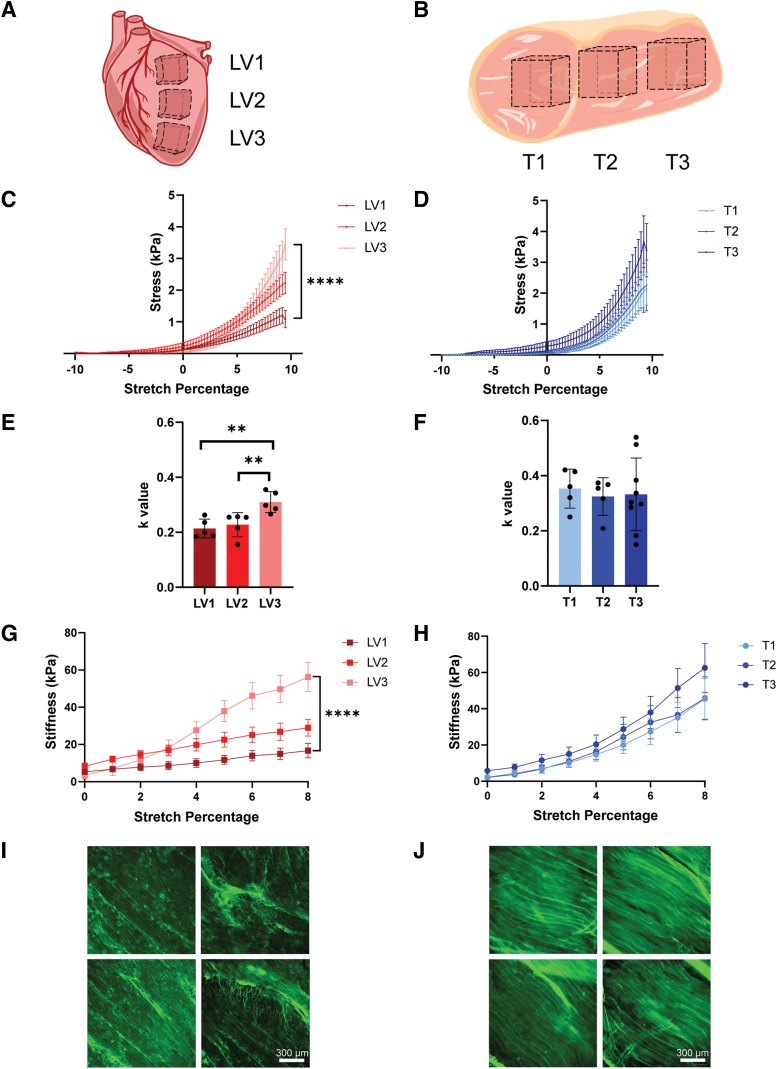
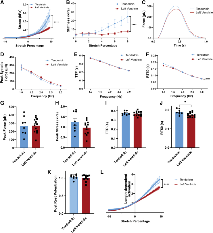
Decellularized porcine myocardium is commonly used as scaffolding for engineered heart tissues (EHTs). However, structural and mechanical heterogeneity in the myocardium complicate production of mechanically consistent tissues. In this study, we evaluate the porcine psoas major muscle (tenderloin) as an alternative scaffold material. Head-to-head comparison of decellularized tenderloin and ventricular scaffolds showed only minor differences in mean biomechanical characteristics, but tenderloin scaffolds were less variable and less dependent on the region of origin than ventricular samples. The active contractile behavior of EHTs made by seeding tenderloin versus ventricular scaffolds with human-induced pluripotent stem cell-derived cardiomyocytes was also comparable, with only minor differences observed. Collectively, the data reveal that the behavior of EHTs produced from decellularized porcine psoas muscle is almost identical to those made from porcine left ventricular myocardium, with the advantages of being more homogeneous, biomechanically consistent, and readily obtainable.

Keywords: cardiomyocyte; decellularized scaffolds; engineered heart tissue; stem cell; tissue engineering.


Conflict of interest statement

S.G.C. has equity ownership in Propria LLC, which has licensed EHT technology used in the research reported in this publication. This arrangement has been reviewed and approved by the Yale University Conflict of Interest Office. The authors declare no additional competing financial interests. J.A.K. provided consulting and collaborative studies with various pharmaceutical companies, but the content of this work is unrelated to the article.

Figures

FIG. 1.
FIG. 1.
EHT production and derivation of analytical measurements. (A) Porcine LV free wall is cut into 150 μm slices, from which scaffolds are laser cut. The scaffolds are decellularized, seeded with iPSC-CMs, cultured, and tested using a custom setup. (B) The TTP and time to 50% relaxation (RT50) are shown on the representative twitch. (C) Passive stress is derived by dividing the force by the cross-sectional area as the tissue is stretched from −10% to 10% stretch. (D) Cross-sectional area of the tissue is taken using OCT. (E) Stiffness is derived from the slope of the stress. TTP, time to peak; iPSC-CM, induced pluripotent stem cell-derived cardiomyocytes; EHT, engineered heart tissue; OCT, optical coherence tomography; LV, left ventricular. Color images are available online.
FIG. 2.
FIG. 2.
Decellularized porcine tenderloin scaffolds are more consistent than LV scaffolds in passive mechanics. (A) Representation of the three different sections taken from porcine LV free wall: LV1 n = 5, LV2 n = 5, and LV3 n = 5. (B) Representation of the three different sections taken from porcine tenderloin (T): T1 n = 5, T2 n = 5, and T3 n = 10. (C) Passive stress mechanics of LV scaffolds being stretched from −10% to 10% (p < 0.0001). (D) Passive stress of T scaffolds stretched from −10% to 10%. (E) LV k-values from the exponential curve fit equation y = Ceka with alpha representing strain (p = 0.0034 for LV1 vs. LV3 and p = 0.0082 for LV2 vs. LV3). (F) Tenderloin passive stress exponential fit k-values. (G) Stiffness for LV scaffolds (p < 0.0001). (H) Stiffness for T scaffolds. (I) Immunohistochemistry (IHC) of LV1 decellularized scaffolds staining for collagen I at 10×. (J) IHC of collagen-stained T1 scaffolds at 10×. Experiments were analyzed with 2-way ANOVA (C, D, F) with mixed effects (G, H) and Tukey’s post hoc comparison (E). **p < 0.01; ****p < 0.0001. Color images are available online.
FIG. 3.
FIG. 3.
Similar passive mechanics and active mechanics, as well as improved Frank-Starling behavior are observed in recellularized tenderloin scaffolds. Both decellularized left ventricle (LV1) and tenderloin (T1) were seeded with PGP1 iPSC-CMs and adult cardiac fibroblasts. All data were from two separate differentiation batches (LV1 n = 12, T1 n = 8). (A) Passive stress of EHTs stretched from −10% to 10% stretch (2-way ANOVA) p < 0.0001). (B) Stiffness as a derivative of passive stress from culture length to 8% stretch (2-way ANOVA p < 0.0001). (C) Representative force twitches of EHT at 1 Hz. (D) Raw peak systolic force from 1 to 3 Hz. (E) TTP force from 1 to 3 Hz. (F) Time to 50% relaxation (RT50) (2-way ANOVA with repeated measures p = 0.0057). (G) Peak systolic force 1 Hz comparison. (H) Peak stress 1 Hz comparison. (I) TTP 1 Hz comparison. (J) RT50 1 Hz comparison (p = 0.0271). (K) Postrest potentiation relationship. (L) Frank-Starling gain from −10% to 10% stretch. Data were analyzed using 2-way ANOVA (A, B, D–F, L) and unpaired two-tailed t test (G–K). *p < 0.05; **p < 0.01; ****p < 0.0001. Color images are available online.
FIG. 4.
FIG. 4.
Mass spectrometry of tenderloin and LV decellularized scaffolds shows similar composition. (A) Summary data of total protein and major ECM proteins in tenderloin and LV decellularized scaffolds. (B) Comparison of collagen composition. (C) Comparison of glycoprotein composition. (D) Comparison of proteoglycan composition. (E) Comparison of other fibers in the ECM. ECM, extracellular matrix. Color images are available online.

References

    1. Jung JP, Hu D, Domian IJ, et al. . An integrated statistical model for enhanced murine cardiomyocyte differentiation via optimized engagement of 3D extracellular matrices. Sci Rep 2015;5:18705; doi: 10.1038/SREP18705 - DOI - PMC - PubMed
    1. Schwan J, Kwaczala AT, Ryan TJ, et al. . Anisotropic engineered heart tissue made from laser-cut decellularized myocardium. Sci Rep 2016;6:32068. - PMC - PubMed
    1. Ott HC, Matthiesen TS, Goh SK, et al. . Perfusion-decellularized matrix: Using nature's platform to engineer a bioartificial heart. Nat Med 2008;14:213–221. - PubMed
    1. Guyette JP, Charest JM, Mills RW, et al. . Bioengineering human myocardium on native extracellular matrix. Circ Res 2016;118:56–72. - PMC - PubMed
    1. Crapo PM, Gilbert TW, Badylak SF. An overview of tissue and whole organ decellularization processes. Biomaterials 2011;32:3233–3243. - PMC - PubMed

Publication types

LinkOut - more resources