. 2023 Jul 14;18(7):e0288412.
doi: 10.1371/journal.pone.0288412. eCollection 2023.

Dynamic susceptibility contrast perfusion-weighted and diffusion-weighted magnetic resonance imaging findings in pilocytic astrocytoma and H3.3 and H3.1 variant diffuse midline glioma, H3K27-altered

Affiliations

Dynamic susceptibility contrast perfusion-weighted and diffusion-weighted magnetic resonance imaging findings in pilocytic astrocytoma and H3.3 and H3.1 variant diffuse midline glioma, H3K27-altered

Ryo Kurokawa et al. PLoS One. .

Abstract

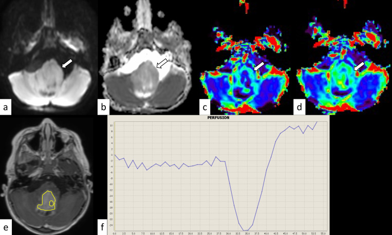
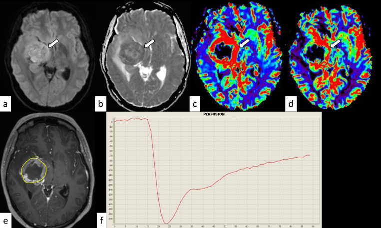
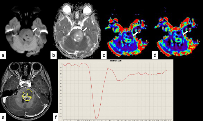
Objective: This study compared the dynamic susceptibility contrast (DSC) magnetic resonance imaging parameters and apparent diffusion coefficient (ADC) between pilocytic astrocytoma (PA) and diffuse midline glioma, H3K27-altered (DMG) variants.

Methods: The normalized relative cerebral blood volume (nrCBV), normalized relative flow (nrCBF), percentile signal recovery (PSR), and normalized mean ADC (nADCmean) of 23 patients with midline PAs (median age, 13 years [range, 1-71 years]; 13 female patients) and 40 patients with DMG (8.5 years [1-35 years]; 19 female patients), including 35 patients with H3.3- and five patients with H3.1-mutant tumors, treated between January 2016 and May 2022 were statistically compared.

Results: DMG had a significantly lower nADCmean (median: 1.48 vs. 1.96; p = 0.00075) and lower PSR (0.97 vs. 1.23, p = 0.13) but higher nrCBV and nrCBF (1.66 vs. 1.17, p = 0.058, respectively, and 1.87 vs. 1.19, p = 0.028, respectively) than PA. The H3.3 variant had a lower nADCmean than the H3.1 variant (1.46 vs. 1.80, p = 0.10).

Conclusion: DMG had lower ADC and PSR and higher rCBV and rCBF than PA. The H3.3 variant had a lower ADC than the H3.1 variant. Recognizing the differences and similarities in the DSC parameters and ADC between these tumors may help presurgical diagnosis.


Conflict of interest statement

The authors have declared that no competing interests exist.

Figures

Fig 1
Fig 1. Pontine pilocytic astrocytoma in a 3-year-old boy.
The mass shows isointensity on diffusion-weighted imaging (a, arrow) and an increased apparent diffusion coefficient (nADCmean = 1.52) (b, arrow). The normalized relative cerebral blood volume and flow are 1.79 and 1.90, respectively (c and d, arrows). The corresponding post-contrast T1-weighted image and region-of-interest in this slice are shown (e). The percentage signal recovery of the mass is 1.50 (f). nADCmean = normalized mean apparent diffusion coefficient.
Fig 2
Fig 2. H3.3 variant diffuse midline glioma, H3K27-altered in a 28-year-old woman.
The mass shows hyperintensity on diffusion-weighted imaging (a, arrow) and a decreased apparent diffusion coefficient (nADCmean is 1.18) (b, arrow). The normalized relative cerebral blood volume and flow are 2.93 and 2.10, respectively (c and d, arrows). The corresponding post-contrast T1-weighted image and region-of-interest in this slice are shown (e). The percentage signal recovery of the mass is 0.63 (f). nADCmean = normalized mean apparent diffusion coefficient.
Fig 3
Fig 3. H3.1 variant diffuse midline glioma, H3K27-altered in a 4-year-old girl.
The mass shows isointensity on diffusion-weighted imaging (a, arrow) and an increased apparent diffusion coefficient (nADCmean = 1.72) (b, arrow). The normalized relative cerebral blood volume and flow are 1.76 and 1.56, respectively (c and d, arrows). The corresponding post-contrast T1-weighted image and region-of-interest in this slice are shown (e). The percentage signal recovery of the mass is 0.78 (f). nADCmean = normalized mean apparent diffusion coefficient.

References

    1. Louis DN, Perry A, Wesseling P, Brat DJ, Cree IA, Figarella-Branger D, et al.. The 2021 WHO Classification of Tumors of the Central Nervous System: a summary. Neuro Oncol. 2021;23: 1231–1251. doi: 10.1093/neuonc/noab106 - DOI - PMC - PubMed
    1. Collins VP, Jones DTW, Giannini C. Pilocytic astrocytoma: pathology, molecular mechanisms and markers. Acta Neuropathol. 2015;129: 775–788. doi: 10.1007/s00401-015-1410-7 - DOI - PMC - PubMed
    1. Salles D, Laviola G, Malinverni AC de M, Stávale JN. Pilocytic astrocytoma: A review of general, clinical, and molecular characteristics. J Child Neurol. 2020;35: 852–858. doi: 10.1177/0883073820937225 - DOI - PubMed
    1. Schulte JD, Buerki RA, Lapointe S, Molinaro AM, Zhang Y, Villanueva-Meyer JE, et al.. Clinical, radiologic, and genetic characteristics of histone H3 K27M-mutant diffuse midline gliomas in adults. Neurooncol Adv. 2020;2: vdaa142. doi: 10.1093/noajnl/vdaa142 - DOI - PMC - PubMed
    1. Findlay IJ, De Iuliis GN, Duchatel RJ, Jackson ER, Vitanza NA, Cain JE, et al.. Pharmaco-proteogenomic profiling of pediatric diffuse midline glioma to inform future treatment strategies. Oncogene. 2022;41: 461–475. doi: 10.1038/s41388-021-02102-y - DOI - PMC - PubMed