. 2023 Jul 14;9(28):eadh1403.
doi: 10.1126/sciadv.adh1403. Epub 2023 Jul 14.

Intensive exercise ameliorates motor and cognitive symptoms in experimental Parkinson's disease restoring striatal synaptic plasticity

Affiliations

Intensive exercise ameliorates motor and cognitive symptoms in experimental Parkinson's disease restoring striatal synaptic plasticity

Gioia Marino et al. Sci Adv. .

Abstract

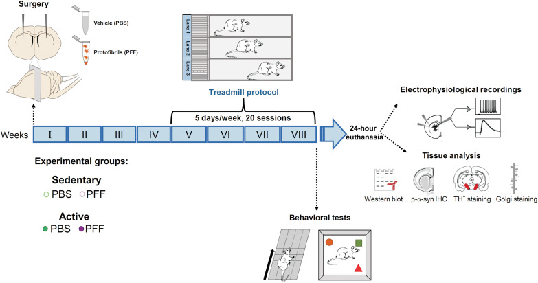
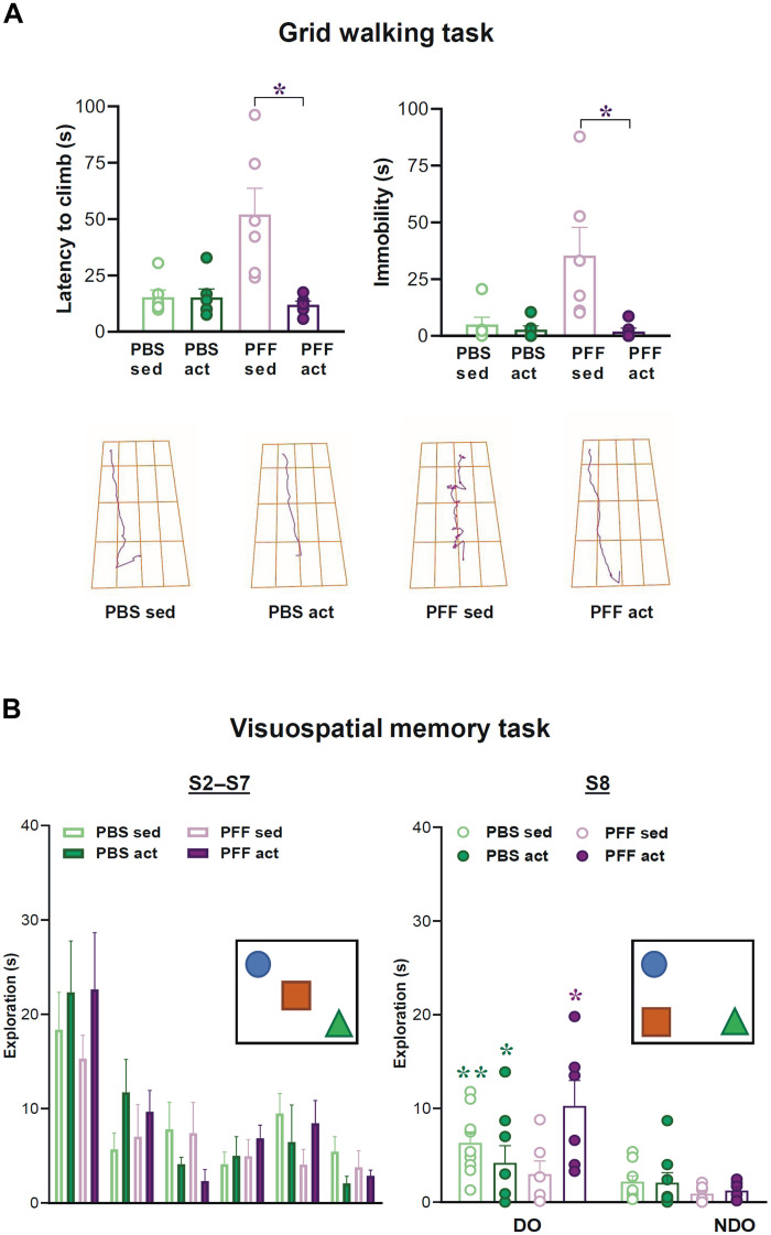
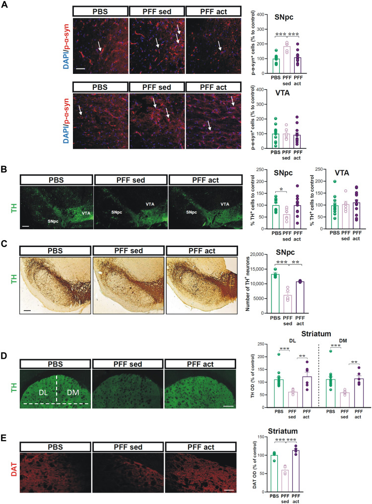
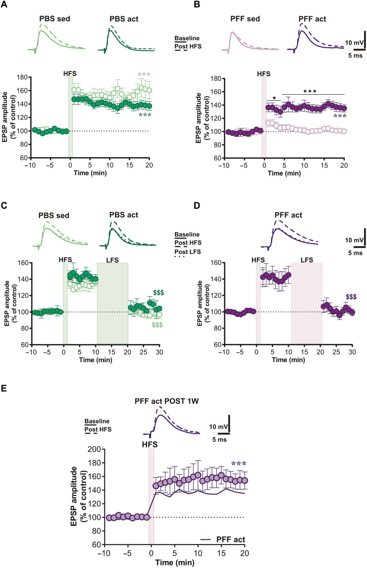
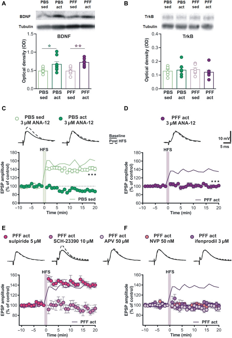
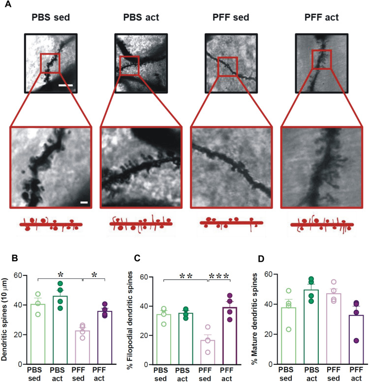
Intensive physical activity improves motor functions in patients with Parkinson's disease (PD) at early stages. However, the mechanisms underlying the beneficial effects of exercise on PD-associated neuronal alterations have not been fully clarified yet. Here, we tested the hypothesis that an intensive treadmill training program rescues alterations in striatal plasticity and early motor and cognitive deficits in rats receiving an intrastriatal injection of alpha-synuclein (α-syn) preformed fibrils. Improved motor control and visuospatial learning in active animals were associated with a recovery of dendritic spine density alterations and a lasting rescue of a physiological corticostriatal long-term potentiation (LTP). Pharmacological analyses of LTP show that modulations of N-methyl-d-aspartate receptors bearing GluN2B subunits and tropomyosin receptor kinase B, the main brain-derived neurotrophic factor receptor, are involved in these beneficial effects. We demonstrate that intensive exercise training has effects on the early plastic alterations induced by α-syn aggregates and reduces the spread of toxic α-syn species to other vulnerable brain areas.


Figures

Fig. 1.
Fig. 1.. Experimental plan.
Schematic representation of the timeline of the experimental procedures and the organization of the experimental groups. Rats were injected with α-syn-PFFs or PBS at 2 to 3 months. Four weeks after the injection, rats were divided into two groups, sedentary and active. The active animals were enrolled in the treadmill protocol for 4 weeks. Sedentary rats were exposed to an experimental apparatus that was switched off. All experimental groups were then subjected to behavioral tests and used for immunofluorescence, electrophysiological, and other morphological experiments. IHC, immunohistochemistry.
Fig. 2.
Fig. 2.. Sensorimotor and visuospatial learning in active and sedentary PFFs-injected parkinsonian animals.
(A) Top: Histograms showing averaged and dot plot values of latency to climb (seconds) and immobility time (seconds) in rats injected with PBS or PFFs and subjected to a 4-week treadmill exercise protocol or a sedentary control condition. Bottom: Representative track plots of the trajectory traveled by rats. *P < 0.05, PFF sed versus all the other groups in the grid test. (B) Left: Histograms illustrating the time (seconds) of object exploration [sessions 2 to 7 (S2 to S7)] during the habituation phase in all experimental groups. Right: Histograms showing the exploration time (seconds) spent exploring a displaced object (DO) and non-displaced objects (NDO) during the session of spatial displacement (S8). Schematic representations of the visuospatial task are reported in the graphs. *P < 0.05, PFF sed versus all the other groups in the grid test. **P < 0.01 PBS sed (DO) versus PBS sed (NDO), *P < 0.05 PBS act (DO) versus PBS act (NDO) and PFF act (DO) versus PFF act (NDO).
Fig. 3.
Fig. 3.. Effects of treadmill on α-syn aggregates diffusion in SNpc and nigrostriatal neurodegeneration.
(A) Left: Representative images at the level of SNpc and VTA showing 4′,6-diamidino-2-phenylindole (DAPI) and p-α-syn immunostaining in PBS- and α-syn-PFF–injected rats, PFF sed, and PFF act, 8 weeks after the injection. White arrows indicate p-α-syn+ dopaminergic neurons. Scale bar, 100 μm. Right: Graphs show the number of p-α-syn+ cells of the SNpc and VTA. ***P < 0.001. (B) Representative images (top) at the level of SNpc and VTA in the different experimental groups. Scale bar, 100 μm. Graphs (bottom) show the number of TH+ neurons, % of cells to control, counted in SNpc and VTA in the different experimental groups. *P < 0.05. (C) Representative images of coronal brain sections showing TH immunoreactivity in the SNpc of the different experimental groups. Scale bar, 200 μm. The graph shows the number of TH+ neurons in the different experimental groups obtained by unbiased stereological analysis. **P < 0.01 and ***P < 0.001. (D) Representative images of coronal brain sections showing TH immunoreactivity in the dorsolateral (DL) and dorsomedial (DM) portion of the striatum in the different experimental groups. Scale bar, 100 μm. The graph shows TH optical density (OD), expressed as a percentage of the control, in both the DL and DM portions of the striatum of the different experimental groups. **P < 0.01 and ***P < 0.001. (E) Representative images of coronal brain sections at the level of the striatum showing DAT immunoreactivity in the different experimental groups. Scale bar, 50 μm. The graph shows DAT OD, expressed as a percentage of the control, in the striatum of the different experimental groups. ***P < 0.001.
Fig. 4.
Fig. 4.. Analysis of corticostriatal synaptic plasticity recorded in striatal neurons of PBS- and PFFs-injected active and sedentary animals.
Time course of excitatory postsynaptic potential (EPSP) amplitude in response to an LTP protocol in SPNs recorded from rats subjected to (A) PBS or (B) PFF intrastriatal injection and enrolled into a treadmill exercise training protocol. Time course of EPSP amplitude changes recorded in SPNs of (C) PBS- and (D) PFF-injected rats in response to the application of HFS, in the absence of magnesium ions, and a subsequent LFS protocol. Scale bars, 5 ms/10 mV. Top: Representative traces of single SPNs recorded from PBS and PFF groups before (solid lines) and after HFS (dashed lines). (E) Time course of EPSP amplitude in response to LTP protocol in SPNs recorded from PFF rats subjected to treadmill exercise training protocol and after 1 week from the last training session. *P < 0.05, ***P < 0.001, and $$$P < 0.001.
Fig. 5.
Fig. 5.. Changes in the protein levels of striatal BDNF and involvement of BDNF-TrkB receptor pathway and GluN2B subunits in the exercise-induced LTP.
(A and B) OD representing the BDNF and TrkB levels in striatal homogenates obtained from brain samples of rats injected with either PBS or PFF and then subjected (act) or not (sed) to treadmill exercise training and evaluated by Western blot. (C and D) Time course of LTP in striatal SPNs from all experimental groups, recorded after 1 hour of incubation with TrkB inhibitor ANA-12 at the concentration of 3 μM. Time course of LTP in striatal SPNs from the PFF act group, recorded during bath application of (E) dopamine receptors (DAR) antagonists (SCH-23390 10 μM and sulpiride 5 μM) and (F) NMDAR antagonists (APV 50 μM, NVP 50 nM, and ifenprodil 3 μM). Scale bars, 5 ms/10 mV. Top: Representative traces of single SPNs recorded from PBS and PFF groups before (solid lines) and after HFS (dashed lines).
Fig. 6.
Fig. 6.. Analysis of the spine density on SPNs of PBS- and PFFs-induced sedentary and active rats.
(A) Representative images of Golgi-stained dendritic segments of a striatal SPN in the different experimental groups at lower (top) and higher (bottom) magnifications. Scale bars, 10 μm (top) and 5 μm (bottom). (B) Histograms of the spine density (n spines per 10 μm) showing the effects of PBS or PFF intrastriatal injection and the subsequent effects of sedentary (sed) condition or treadmill (act) protocol on SPN spine density. *P < 0.05. (C) Histogram showing the quantification of filopodial spines expressed as a percentage of the total number of spines displaying the effects of the different treatments on the immature spines. **P < 0.01 and ***P < 0.001. (D) Histogram showing the amount of mature spines expressed as a percentage of the total number of spines. No substantial effects of PBS or PFF intrastriatal injection and of sedentary (sed) or treadmill (act) protocol on mature spines were detected.

References

    1. W. Poewe, K. Seppi, C. M. Tanner, G. M. Halliday, P. Brundin, J. Volkmann, A.-E. Schrag, A. E. Lang, Parkinson disease. Nat. Rev. Dis. Primers 3, 17013 (2017). - PubMed
    1. M. Schenkman, C. G. Moore, W. M. Kohrt, D. A. Hall, A. Delitto, C. L. Comella, D. A. Josbeno, C. L. Christiansen, B. D. Berman, B. M. Kluger, E. L. Melanson, S. Jain, J. A. Robichaud, C. Poon, D. M. Corcos, Effect of high-intensity treadmill exercise on motor symptoms in patients with de novo parkinson disease: A phase 2 randomized clinical trial. JAMA Neurol. 75, 219–226 (2018). - PMC - PubMed
    1. M. A. Sacheli, J. L. Neva, B. Lakhani, D. K. Murray, N. Vafai, E. Shahinfard, C. English, S. McCormick, K. Dinelle, N. Neilson, J. McKenzie, M. Schulzer, D. C. McKenzie, S. Appel-Cresswell, M. J. McKeown, L. A. Boyd, V. Sossi, A. J. Stoessl, Exercise increases caudate dopamine release and ventral striatal activation in Parkinson's disease. Mov. Disord. 34, 1891–1900 (2019). - PubMed
    1. N. M. van der Kolk, N. M. de Vries, R. P. C. Kessels, H. Joosten, A. H. Zwinderman, B. Post, B. R. Bloem, Effectiveness of home-based and remotely supervised aerobic exercise in Parkinson's disease: A double-blind, randomised controlled trial. Lancet Neurol. 18, 998–1008 (2019). - PubMed
    1. M. E. Johansson, I. G. M. Cameron, N. M. Van der Kolk, N. M. de Vries, E. Klimars, I. Toni, B. R. Bloem, R. C. Helmich, Aerobic exercise alters brain function and structure in parkinson's disease: A randomized controlled trial. Ann. Neurol. 91, 203–216 (2022). - PMC - PubMed