. 2023 Jul 14;9(28):eadh2264.
doi: 10.1126/sciadv.adh2264. Epub 2023 Jul 14.

Lrp1 is essential for lethal Rift Valley fever hepatic disease in mice

Affiliations

Lrp1 is essential for lethal Rift Valley fever hepatic disease in mice

Madeline M Schwarz et al. Sci Adv. .

Abstract

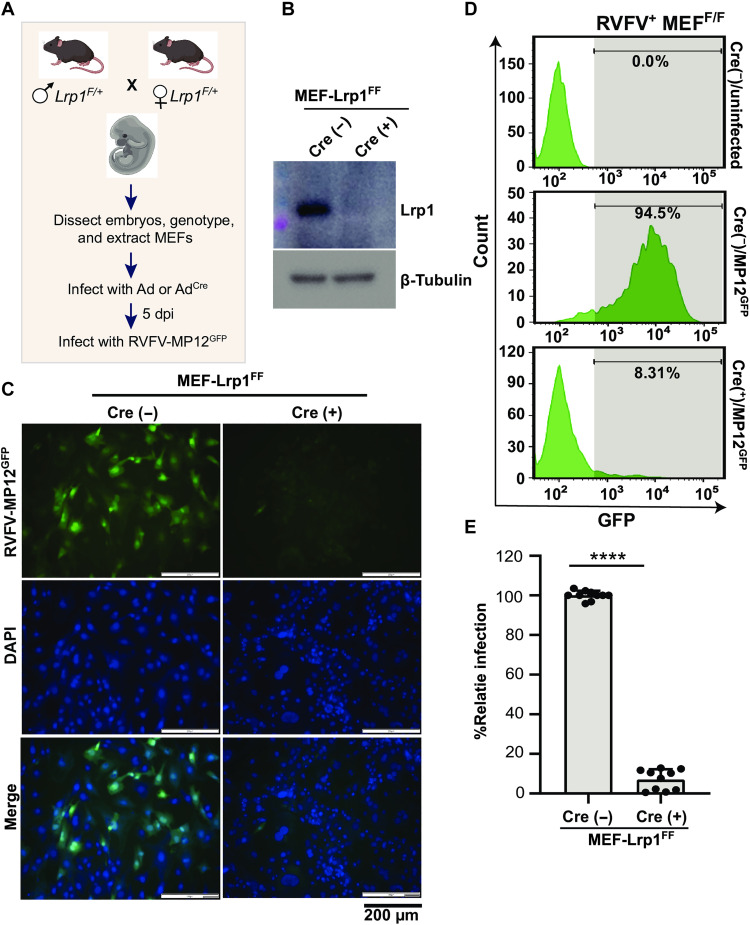
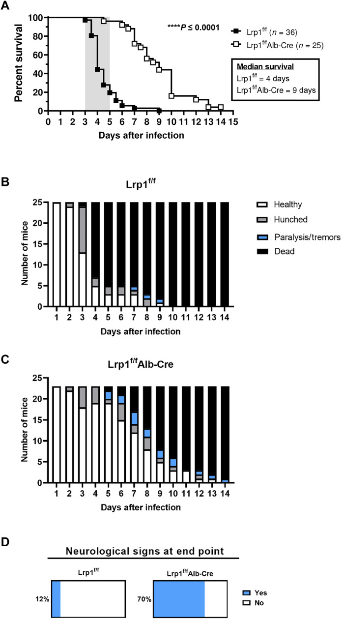
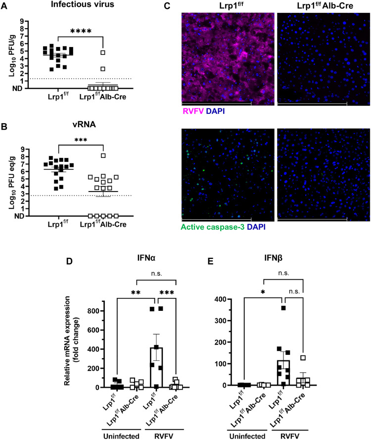
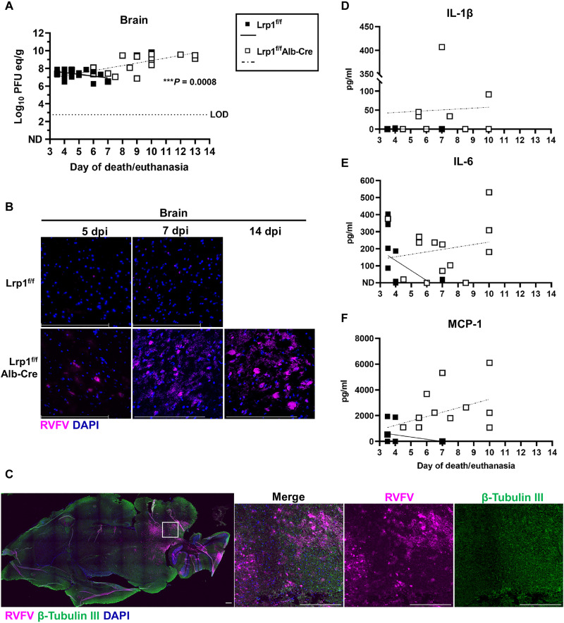
Rift Valley fever virus (RVFV) is an emerging arbovirus found in Africa. While RVFV is pantropic and infects many cells and tissues, viral replication and necrosis within the liver play a critical role in mediating severe disease. The low-density lipoprotein receptor-related protein 1 (Lrp1) is a recently identified host factor for cellular entry and infection by RVFV. The biological significance of Lrp1, including its role in hepatic disease in vivo, however, remains to be determined. Because Lrp1 has a high expression level in hepatocytes, we developed a mouse model in which Lrp1 is specifically deleted in hepatocytes to test how the absence of liver Lrp1 expression affects RVF pathogenesis. Mice lacking Lrp1 expression in hepatocytes showed minimal RVFV replication in the liver, longer time to death, and altered clinical signs toward neurological disease. In contrast, RVFV infection levels in other tissues showed no difference between the two genotypes. Therefore, Lrp1 is essential for RVF hepatic disease in mice.


Figures

Fig. 1.
Fig. 1.. Lrp1-depleted MEFs are resistant to RVFV infection.
(A) Schematics showing the generation of mouse embryonic fibroblasts (MEFs) from embryonic day (E) 14.5 embryos obtained by timed mating of Lrp1f/+ mice. dpi, days postinfection. (B) Western blot of Lrp1f/f MEFs infected with adenovirus expressing an empty adenovirus vector (Ad) and the cre recombinase enzyme (Adcre). The blot was probed for lipoprotein receptor–related protein 1 (Lrp1) and β-tubulin. Lrp1-sufficient and Lrp1-deficient MEFs were infected with Rift Valley fever virus (RVFV) MP12GFP at a multiplicity of infection (MOI) of 2 for 16 hours. (C) Fluorescence images show green fluorescent protein (GFP), DAPI (4′,6-diamidino-2-phenylindole), and merged panels. Images were taken at ×20 magnification (scale bars, 200 μm). (D and E) Representative flow cytometry histograms and corresponding analysis of flow cytometry histogram data from MEF infections. Experiment was performed five times in duplicates. Student’s t test was used to determine significance; ****P < 0.0001.
Fig. 2.
Fig. 2.. Elimination of Lrp1 in hepatocytes significantly delays time to death after RVFV infection.
(A) Lrp1f/f and Lrp1f/fAlb-Cre mice were infected with 20 plaque-forming units (PFUs) of RVFV ZH501 by footpad and monitored for survival (n = 61 animals, data were derived from three experiments). Clinical signs of (B) Lrp1f/f mice and (C) Lrp1f/fAlb-Cre mice highlight the frequency of neurological symptoms (note that mice found deceased are not included in this scoring). (D) Percentages of mice across three studies that experienced neurological signs of disease including tremors and paralysis (light blue) on the day of euthanasia in Lrp1f/f and Lrp1f/fAlb-Cre mice. Mice that were found deceased could not be clinically scored and were not included in analysis in (D). Kaplan-Meier survival analysis was completed to assess for significant differences between genotypes. ****P < 0.0001.
Fig. 3.
Fig. 3.. Lrp1 KO in hepatocytes leads to a significant reduction in viral burden in the liver at 3 dpi.
(A) Lrp1f/fAlb-Cre mice (n = 16) have significantly less (A) infectious virus and (B) viral RNA (vRNA) in the liver at 3 dpi than Lrp1f/f controls (n = 15). (C) RVFV nucleoprotein (N) (magenta) and active caspase-3 (green) staining in the liver at 3 dpi in Lrp1f/f and Lrp1f/fAlb-Cre livers (40×). (D) Interferon α (IFNα) and (E) IFNβ relative mRNA expression in the liver of uninfected and RVFV-infected Lrp1f/f and Lrp1f/fAlb-Cre mice at 3 dpi. Bars represent SEM. Statistics were determined by an unpaired t test or a one-way analysis of variance (ANOVA). *P < 0.05; **P < 0.01; ***P < 0.001; ****P < 0.0001. n.s., not significant. Scale bars, 250 μm. Dotted lines indicate the limit of detection.
Fig. 4.
Fig. 4.. Lrp1 KO in hepatocytes reduces RVFV in the liver at time of euthanasia.
(A) vRNA titers in the liver on the day of euthanasia show a significant negative correlation in Lrp1f/fAlb-Cre mice from 3 to 14 dpi. LOD, limit of assay detection. (B) Gross pathology of liver tissue obtained from the indicated genotypes at 5 dpi. DOA, deceased on arrival. (C) RVFV N antigen staining in liver tissue of Lrp1f/f mice compared to Lrp1f/fAlb-Cre mice at 5 and 7 dpi (40×). (D) Interleukin-1β (IL-1β), (E) IL-6, and (F) MCP-1 protein levels in the liver tissue homogenate on the day of euthanasia from 3 to 14 dpi. Scale bars, 250 μm. A linear regression was used to determine whether the slope was significantly nonzero. **P < 0.01; ***P < 0.001.
Fig. 5.
Fig. 5.. Mice with Lrp1 KO in hepatocytes succumb to RVF neurological disease.
(A) vRNA titers in the brain at euthanasia show a significant positive correlation in Lrp1f/fAlb-Cre mice from 3 to 14 dpi. (B) RVFV staining in the brain tissue of Lrp1f/fAlb-Cre mice compared to Lrp1f/f mice from 5, 7, and 14 dpi (40×). (C) Brain tissue from a Lrp1f/fAlb-cre mice at 12 dpi stained for RVFV N (magenta), β-tubulin III (green), and DAPI (blue; 10×). (D) IL-1β, (E) IL-6, and (F) MCP-1 protein in the brain tissue at euthanasia from 3 to 14 dpi. Scale bars, 250 μm. A linear regression was used to determine whether the slope was significantly nonzero. **P < 0.01.

References

    1. M. Kwaśnik, W. Rożek, J. Rola, Rift valley fever–A growing threat to humans and animals. J. Vet. Res. 65, 7–14 (2021). - PMC - PubMed
    1. R. Métras, W. J. Edmunds, C. Youssouffi, L. Dommergues, G. Fournié, A. Camacho, S. Funk, E. Cardinale, G. le Godais, S. Combo, L. Filleul, H. Youssouf, M. Subiros, Estimation of rift valley fever virus spillover to humans during the Mayotte 2018-2019 epidemic. Proc. Natl. Acad. Sci. U.S.A. 117, 24567–24574 (2020). - PMC - PubMed
    1. L. W. Laughlin, J. M. Meegan, L. J. Strausbaugh, D. M. Morens, R. H. Watten, Epidemic rift valley fever in Egypt: Observations of the spectrum of human illness. Trans. R. Soc. Trop. Med. Hyg. 73, 630–633 (1979). - PubMed
    1. T. Ikegami, S. Makino, The pathogenesis of rift valley fever. Viruses 3, 493–519 (2011). - PMC - PubMed
    1. M. Al-Hazmi, E. A. Ayoola, M. Abdurahman, S. Banzal, J. Ashraf, A. El-Bushra, A. Hazmi, M. Abdullah, H. Abbo, A. Elamin, E.-T. Al-Sammani, M. Gadour, C. Menon, M. Hamza, I. Rahim, M. Hafez, M. Jambavalikar, H. Arishi, A. Aqeel, Epidemic rift valley fever in Saudi Arabia: A clinical study of severe illness in humans. Clin. Infect. Dis. 36, 245–252 (2003). - PubMed

Substances