. 2023 Dec 1;278(6):e1313-e1326.
doi: 10.1097/SLA.0000000000006005. Epub 2023 Jul 14.

Mitochondria Transplantation Mitigates Damage in an In Vitro Model of Renal Tubular Injury and in an Ex Vivo Model of DCD Renal Transplantation

Affiliations

Mitochondria Transplantation Mitigates Damage in an In Vitro Model of Renal Tubular Injury and in an Ex Vivo Model of DCD Renal Transplantation

Andrea Rossi et al. Ann Surg. .

Abstract

Objectives: To test whether mitochondrial transplantation (MITO) mitigates damage in 2 models of acute kidney injury (AKI).

Background: MITO is a process where exogenous isolated mitochondria are taken up by cells. As virtually any morbid clinical condition is characterized by mitochondrial distress, MITO may find a role as a treatment modality in numerous clinical scenarios including AKI.

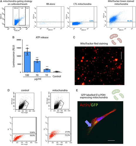
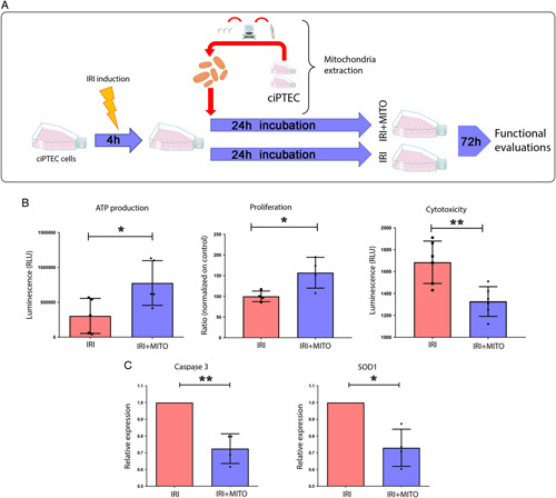
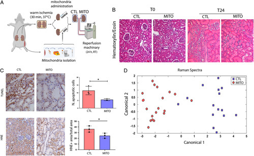
Methods: For the in vitro experiments, human proximal tubular cells were damaged and then treated with mitochondria or placebo. For the ex vivo experiments, we developed a non-survival ex vivo porcine model mimicking the donation after cardiac death renal transplantation scenario. One kidney was treated with mitochondria, although the mate organ received placebo, before being perfused at room temperature for 24 hours. Perfusate samples were collected at different time points and analyzed with Raman spectroscopy. Biopsies taken at baseline and 24 hours were analyzed with standard pathology, immunohistochemistry, and RNA sequencing analysis.

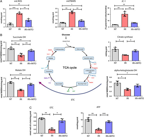
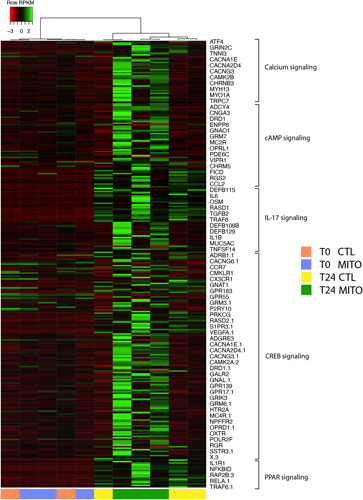
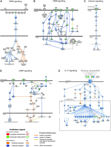
Results: In vitro, cells treated with MITO showed higher proliferative capacity and adenosine 5'-triphosphate production, preservation of physiological polarization of the organelles and lower toxicity and reactive oxygen species production. Ex vivo, kidneys treated with MITO shed fewer molecular species, indicating stability. In these kidneys, pathology showed less damage whereas RNAseq analysis showed modulation of genes and pathways most consistent with mitochondrial biogenesis and energy metabolism and downregulation of genes involved in neutrophil recruitment, including IL1A, CXCL8, and PIK3R1.

Conclusions: MITO mitigates AKI both in vitro and ex vivo.


Conflict of interest statement

J.R. and R.S.S. are co-founders of Rametrix Technologies, on the board of which J.R. serves as President and CEO, whereas R.S.S. as Chief Technology Officer. The remaining authors report no conflicts of interest.

Figures

FIGURE 1
FIGURE 1
Isolation and internalization of viable mitochondria. A, Isolated mitochondria were suspended in resuspension buffer (RB) and stained with MitoTracker Green FM 200 nM. Calibrated beads were used to gate events of 0.5 to 3 μm. A representative cytofluorimetric analysis shows the absence of fluorescence in RB with MitoTracker Green or in unstained mitochondria. Fluorescence intensity of MitoTracker Green labeled mitochondria, reaching >90% events, was observed among the 0.5 to 1 μm gates. B, Adenosine 5′-triphosphate (ATP) production by isolated mitochondria resuspended in RB at different concentrations (100–10 µg/ml), showed a mitochondrial dose dependent increase in ATP concentration. Control: RB alone. n=3. One-way analysis of variance (ANOVA) with Dunnett multicomparison test was performed: ***P < 0.001 versus Control. C, A representative image of isolated viable mitochondria labeled with MitoTracker red. Scalebar: 5 μm. D, Representative cytofluorimetric analysis shows the acquisition of MitoTracker red labeled mitochondria in MITO human conditionally immortalized proximal tubular cell (ciPTECs). E, A representative image of mitochondria expressing the GFP-labeled E1α pyruvate dehydrogenase after MITO shows the presence of GFP-labeled mitochondria within ciPTECs. Actin is labeled in red. Scalebar: 5 μm. CTL indicates control.
FIGURE 2
FIGURE 2
Mitochondrial transplantation (MITO) reduces reactive oxygen species and mitigates mitochondrial damage in IRI. A, Human conditionally immortalized proximal tubular cell (ciPTEC) were subjected to IRI and co-incubated without (IRI) or with mitochondria (20 µg/ml resuspension buffer, IRI+MITO) isolated from healthy ciPTECs. After 24 hours, cells were washed 3 times, cultured for further for 72 hours, and mitochondria isolated for functional evaluation. B, IRI damaged ciPTECs were compared with IRI damaged ciPTECs treated with MITO. MITO promoted ATP production, proliferation and reduced cytotoxicity compared with IRI damaged cells. C, Expression of apoptotic genes, Caspase-3 and superoxide dismutase (SOD1) was assessed in IRI ciPTECs and compared with IRI+MITO ciPTECs. qRT-PCR results showed a significant decrease in apoptosis in IRI+MITO ciPTECs (mean ± SD normalized to GAPDH and expressed relative to untreated IRI ciPTECs). Data are mean ±SD of 4 different experiments, Student t test was performed: *P< 0.05, **P < 0.01 vs IRI). ATP indicates adenosine 5′-triphosphate.
FIGURE 3
FIGURE 3
MITO improves mitochondrial metabolism after IRI. Normal healthy human conditionally immortalized proximal tubular cells (ciPTECs) (NT) or IRI damaged ciPTECs were treated without (IRI) or with mitochondrial transplantation (MITO) (20 µg/ml, IRI+MITO). A, MITO normalized reactive oxygen species (ROS) levels, mitochondrial thiobarbituric reactive substances and JC-1 membrane potential altered by IRI. B, Key tricarboxylic acid (TCA) cycle enzyme [succinate dehydrogenase (DH), citrate synthase, malate DH] activities in IRI ciPTECs were significantly increased by MITO (IRI+MITO). C, Electron transport chain (ETC) and adenosine 5′-triphosphate (ATP) concentrations in IRI ciPTECs were significantly increased with MITO treatment (IRI+MITO). Data are mean ±SD of 4 different experiments, one-way analysis of variance (ANOVA) with Tukey multicomparison test was performed: *P< 0.05, **P < 0.01, ***P < 0.001.
FIGURE 4
FIGURE 4
MITO improves IRI in a model of donation after cardiac death (DCD) renal transplant. A, An ex vivo model of DCD kidneys was used to test the hypothesis that mitochondrial transplantation (MITO) improves the pathophysiology associated with IRI. Kidneys were subjected to warm ischemia for 30 minutes, followed by injection with vehicle (CTL) or mitochondria isolated from 1g of autologous psoas muscle (MITO). After 30 min, kidneys were perfused for 24 hours. (n=4 animals, 5 kidneys (one CTL discarded), 2 experimental groups, CTL and MITO). B, The presence of macroscopic damage in control (CTL) and mitochondrial treated (MITO) kidneys was shown by hematoxylin and eosin staining. C, thymidine deoxyribose-mediated deoxy-UTP nick end labeling (TUNEL) assay and 4-hydroxy-2-nonenal (HNE) showed a decrease of apoptotic cells and lipid peroxidation, respectively, in MITO kidneys (representative images) that was confirmed by quantification. Student t test was performed: *P < 0.05. D, Raman spectra, collected at sequential timepoints from control and MITO kidneys were baseline normalized and then analyzed using principal component analysis and discriminate analysis of principal components. The canonical plot matrix shows differences in the molecular composition of perfusate from control and MITO kidneys appearing as definitive separated clusters. MITO treated kidneys shed fewer molecules/molecular species over 24 hours than CTL kidneys, indicating greater stability/viability [shown statistically by calculation of Total Spectral Distance vs Analytical Standard (Surine) using the RametrixTM Toolboxes]. CTL indicates control.
FIGURE 5
FIGURE 5
Mitochondrial transplantation (MITO) modulates mitochondrial biogenesis and mitochondrial metabolism pathways in a donation after cardiac death kidney model. Pathway analysis was performed using Ingenuity Pathway Analysis software (IPA, Qiagen). Selection of the pathways was based on the significant modulation of transcripts between MITO T24 versus control (CTL) T24 (fold change >1.5 or < −1.5; P < 0.05). The hierarchical clustering heatmap shows calcium, cAMP, IL-17, CREB, and PPAR signaling pathway specific transcript modulation in control (CTL) and treated (MITO) at T0 and T24. Values for differentially expressed genes in these 5 pathways are reported in Suppl. Table 5, Supplemental Digital Content 1, http://links.lww.com/SLA/E728.
FIGURE 6
FIGURE 6
Mitochondrial transplantation (MITO) increases intracellular signaling pathway genes in a donation after cardiac death kidney model. Pathway analysis for transcript modulations after 24 hour perfusion between treatment (MITO T24) versus control (CTRL T24) (fold change >1.5 or < −1.5; P <0.05) was performed using Ingenuity Pathway Analysis software (IPA, Qiagen). Increased expression of signaling pathway genes was observed, including G proteins/adenylyl cyclases/cAMP signaling mediators, MAPK/ERK and PI3K/AKT genes, and increased levels of ELK1 and CREB transcription factors. Results show the interaction of common activated signaling pathway genes after MITO. Upregulated genes in dataset are represented in blue.

References

    1. Brennan C, Husain SA, King KL, et al. . A donor utilization index to assess the utilization and discard of deceased donor kidneys perceived as high risk. Clin J Am Soc Nephrol. 2019;14:1634–1641. - PMC - PubMed
    1. Orlando G, Soker S, Stratta RJ, et al. . Will regenerative medicine replace transplantation? Cold Spring Harb Perspect Med. 2013;3:a015693. - PMC - PubMed
    1. Rao PS, Ojo A. The alphabet soup of kidney transplantation: SCD, DCD, ECD--fundamentals for the practicing nephrologist. Clin J Am Soc Nephrol. 2009;4:1827–1831. - PubMed
    1. Gill J, Rose C, Lesage J, et al. . Use and outcomes of kidneys from donation after circulatory death donors in the United States. J Am Soc Nephrol. 2017;28:3647–3657. - PMC - PubMed
    1. Tapiawala SN, Tinckam KJ, Cardella CJ, et al. . Delayed graft function and the risk for death with a functioning graft. J Am Soc Nephrol. 2010;21:153–161. - PMC - PubMed

Publication types