. 2023 Oct 2;22(10):1204-1214.
doi: 10.1158/1535-7163.MCT-23-0221.

Targeting Solid Cancers with a Cancer-Specific Monoclonal Antibody to Surface Expressed Aberrantly O-glycosylated Proteins

Affiliations

Targeting Solid Cancers with a Cancer-Specific Monoclonal Antibody to Surface Expressed Aberrantly O-glycosylated Proteins

Mikkel K M Aasted et al. Mol Cancer Ther. .

Abstract

The lack of antibodies with sufficient cancer selectivity is currently limiting the treatment of solid tumors by immunotherapies. Most current immunotherapeutic targets are tumor-associated antigens that are also found in healthy tissues and often do not display sufficient cancer selectivity to be used as targets for potent antibody-based immunotherapeutic treatments, such as chimeric antigen receptor (CAR) T cells. Many solid tumors, however, display aberrant glycosylation that results in expression of tumor-associated carbohydrate antigens that are distinct from healthy tissues. Targeting aberrantly glycosylated glycopeptide epitopes within existing or novel glycoprotein targets may provide the cancer selectivity needed for immunotherapy of solid tumors. However, to date only a few such glycopeptide epitopes have been targeted. Here, we used O-glycoproteomics data from multiple cell lines to identify a glycopeptide epitope in CD44v6, a cancer-associated CD44 isoform, and developed a cancer-specific mAb, 4C8, through a glycopeptide immunization strategy. 4C8 selectively binds to Tn-glycosylated CD44v6 in a site-specific manner with low nanomolar affinity. 4C8 was shown to be highly cancer specific by IHC of sections from multiple healthy and cancerous tissues. 4C8 CAR T cells demonstrated target-specific cytotoxicity in vitro and significant tumor regression and increased survival in vivo. Importantly, 4C8 CAR T cells were able to selectively kill target cells in a mixed organotypic skin cancer model having abundant CD44v6 expression without affecting healthy keratinocytes, indicating tolerability and safety.


Figures

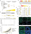
Figure 1. mAb 4C8 demonstrates high specificity toward CD44v6-Tn. A, Schematic illustration demonstrating how increased cancer selectivity can be achieved by targeting cancer-associated glycans within cancer-associated domains of CD44. B, Structure of CD44, highlighting the variable region with all its known O-glycosylation sites, and then the sequence of the cancer-associated CD44v6 domain likewise with O-glycosylation sites highlighted and annotated for which cells lines they have been identified in. C, Variable concentrations of 4C8 were used to perform ELISA against non-glycosylated and Tn-glycosylated CD44v6 and unrelated Tn-glycosylated glycopeptides. D, Affinities for various Tn-glycosylated glycopeptides, including CD44v6 and naked CD44v6 peptide as measured by Octet. E, Various wild-type and COSMCKO cells were stained for flow cytometry using 4C8 and antibodies recognizing Tn-antigen or CD44v6 as controls. Graphs show histograms of cell populations stained with the annotated antibodies. F, 4C8 was used to stain wild-type and COSMCKO HaCaT cells for immunofluorescence microscopy. In addition, cells were stained for Tn expression and non-glycosylated CD44v6. Green, CD44v6, Tn, or 4C8, blue, DAPI. G, 4C8 was used to stain normal human skin by immunofluorescence. Sections were also stained for non-glycosylated CD44v6. Green, CD44v6 or 4C8, blue, DAPI.
Figure 1.
mAb 4C8 demonstrates high specificity toward CD44v6-Tn. A, Schematic illustration demonstrating how increased cancer selectivity can be achieved by targeting cancer-associated glycans within cancer-associated domains of CD44. B, Structure of CD44, highlighting the variable region with all its known O-glycosylation sites, and then the sequence of the cancer-associated CD44v6 domain likewise with O-glycosylation sites highlighted and annotated for which cells lines they have been identified in. C, Variable concentrations of 4C8 were used to perform ELISA against non-glycosylated and Tn-glycosylated CD44v6 and unrelated Tn-glycosylated glycopeptides. D, Affinities for various Tn-glycosylated glycopeptides, including CD44v6 and naked CD44v6 peptide as measured by Octet. E, Various wild-type and COSMCKO cells were stained for flow cytometry using 4C8 and antibodies recognizing Tn-antigen or CD44v6 as controls. Graphs show histograms of cell populations stained with the annotated antibodies. F, 4C8 was used to stain wild-type and COSMCKO HaCaT cells for immunofluorescence microscopy. In addition, cells were stained for Tn expression and non-glycosylated CD44v6. Green, CD44v6, Tn, or 4C8, blue, DAPI. G, 4C8 was used to stain normal human skin by immunofluorescence. Sections were also stained for non-glycosylated CD44v6. Green, CD44v6 or 4C8, blue, DAPI.
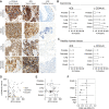
Figure 2. 4C8 selectively stain tumor tissues from several cancers. A, 4C8 antibody, anti-CD44 antibody, and IgG isotype control antibody were used to stain tissue microarrays from pancreatic, breast, prostate, colon, and lung carcinomas. Sections show representative stainings of tumor tissue with anti-CD44v6, 4C8, and adjacent normal tissue with 4C8. B and C, Graphs illustrating the percentage of carcinoma and healthy tissue sections of each tissue type staining positive with 4C8 and non-glycosylated CD44v6. D, Dotplot plotting H-scores of 4C8 staining against CD44v6 staining, individual datapoints colored on the basis of scoring as positive for CD44v6 only (gray), 4C8 only (yellow), or both (black). E, Boxplots showing distribution of 4C8 H-scores within positively scored tumors. F, Odds ratios and 95% confidence intervals for positive 4C8 staining. For cancer grade, odds ratios were calculated compared with grade 1 cancers and were controlled for sex and age. For the cancer stage, odds ratios were calculated compared with stage 1 and were controlled for sex and age. For sex, odds ratios were calculated comparing male with female and were controlled for the grade, stage, and age. For age, odds ratios were controlled for the grade, stage, and sex. *, P < 0.05; **, P < 0.01.
Figure 2.
4C8 selectively stain tumor tissues from several cancers. A, 4C8 antibody, anti-CD44 antibody, and IgG isotype control antibody were used to stain tissue microarrays from pancreatic, breast, prostate, colon, and lung carcinomas. Sections show representative stainings of tumor tissue with anti-CD44v6, 4C8, and adjacent normal tissue with 4C8. B and C, Graphs illustrating the percentage of carcinoma and healthy tissue sections of each tissue type staining positive with 4C8 and non-glycosylated CD44v6. D, Dotplot plotting H-scores of 4C8 staining against CD44v6 staining, individual datapoints colored on the basis of scoring as positive for CD44v6 only (gray), 4C8 only (yellow), or both (black). E, Boxplots showing distribution of 4C8 H-scores within positively scored tumors. F, Odds ratios and 95% confidence intervals for positive 4C8 staining. For cancer grade, odds ratios were calculated compared with grade 1 cancers and were controlled for sex and age. For the cancer stage, odds ratios were calculated compared with stage 1 and were controlled for sex and age. For sex, odds ratios were calculated comparing male with female and were controlled for the grade, stage, and age. For age, odds ratios were controlled for the grade, stage, and sex. *, P < 0.05; **, P < 0.01.
Figure 3. 4C8 CAR T cells selectively kill Tn-expressing cancer cells in vitro. A, Illustration of the construct design of the 4C8CARs: A single scFv domain derived from 4C8 was joined via the hinge region of CD8 to the transmembrane and intracellular signaling domain of CD28, and finally the intracellular signaling domain of CD3ζ. B, Histograms of flow cytometry data showing 4C8 CAR-positive cell populations in non-transduced (NTD) or transduced T cells. C, NTD, 4C8 CAR T cells, or CD19 CAR T cells were placed in co-cultures with various target cells, and the target cell viability and the production of IFNγ was measured.
Figure 3.
4C8 CAR T cells selectively kill Tn-expressing cancer cells in vitro.A, Illustration of the construct design of the 4C8CARs: A single scFv domain derived from 4C8 was joined via the hinge region of CD8 to the transmembrane and intracellular signaling domain of CD28, and finally the intracellular signaling domain of CD3ζ. B, Histograms of flow cytometry data showing 4C8 CAR-positive cell populations in non-transduced (NTD) or transduced T cells. C, NTD, 4C8 CAR T cells, or CD19 CAR T cells were placed in co-cultures with various target cells, and the target cell viability and the production of IFNγ was measured.
Figure 4. Jurkat leukemia controlled in vivo by 4C8 CAR T cells. A, Luminescence images of mice inoculated with Jurkat cells and treated with NTD, CD19, or 4C8 CAR T cells (n = 10). B, Quantification of luminescence signal from mice. C, Survival curve of mice in days after T-cell infusion.
Figure 4.
Jurkat leukemia controlled in vivo by 4C8 CAR T cells. A, Luminescence images of mice inoculated with Jurkat cells and treated with NTD, CD19, or 4C8 CAR T cells (n = 10). B, Quantification of luminescence signal from mice. C, Survival curve of mice in days after T-cell infusion.
Figure 5. 4C8 CAR T cells show selective killing in organotypic skin tumor model. A, Illustration of the experimental setup and production of organotypic skin tumor models. B, In vitro cytotoxicity of 4C8 CAR T cells against wild-type and COSMC-deficient N/TERT-1 cells. C, Immunofluorescence micrographs of sectioned organotypic models stained for Tn-antigen (green) and CD44 (red) or keratin 10 (green) and keratin 5 (red). D, Graphs quantifying the percentage of Tn-positive hRasKI Core 1KO keratinocytes in the skin model (n = 5). *, P < 0.05; **, P < 0.01.
Figure 5.
4C8 CAR T cells show selective killing in organotypic skin tumor model. A, Illustration of the experimental setup and production of organotypic skin tumor models. B,In vitro cytotoxicity of 4C8 CAR T cells against wild-type and COSMC-deficient N/TERT-1 cells. C, Immunofluorescence micrographs of sectioned organotypic models stained for Tn-antigen (green) and CD44 (red) or keratin 10 (green) and keratin 5 (red). D, Graphs quantifying the percentage of Tn-positive hRasKI Core 1KO keratinocytes in the skin model (n = 5). *, P < 0.05; **, P < 0.01.

References

    1. Kochenderfer JN, Wilson WH, Janik JE, Dudley ME, Stetler-Stevenson M, Feldman SA, et al. . Eradication of B-lineage cells and regression of lymphoma in a patient treated with autologous T cells genetically engineered to recognize CD19. Blood 2010;116:4099–102. - PMC - PubMed
    1. Kalos M, Levine BL, Porter DL, Katz S, Grupp SA, Bagg A, et al. . T cells with chimeric antigen receptors have potent antitumor effects and can establish memory in patients with advanced leukemia. Sci Transl Med 2011;3:95ra73. - PMC - PubMed
    1. Porter DL, Levine BL, Kalos M, Bagg A, June CH. Chimeric antigen receptor–modified T cells in chronic lymphoid leukemia. N Engl J Med 2011;365:725–33. - PMC - PubMed
    1. Brentjens RJ, Davila ML, Riviere I, Park J, Wang X, Cowell LG, et al. . CD19-targeted T cells rapidly induce molecular remissions in adults with chemotherapy-refractory acute lymphoblastic leukemia. Sci Transl Med 2013;5:177ra38. - PMC - PubMed
    1. Grupp SA, Kalos M, Barrett D, Aplenc R, Porter DL, Rheingold SR, et al. . Chimeric antigen receptor–modified T cells for acute lymphoid leukemia. N Engl J Med 2013;368:1509–18. - PMC - PubMed

Publication types