. 2023 Aug;120(2):298-306.
doi: 10.1111/mmi.15123. Epub 2023 Jul 14.

Escherichia coli DNA repair helicase Lhr is also a uracil-DNA glycosylase

Affiliations

Escherichia coli DNA repair helicase Lhr is also a uracil-DNA glycosylase

Ryan J Buckley et al. Mol Microbiol. 2023 Aug.

Abstract

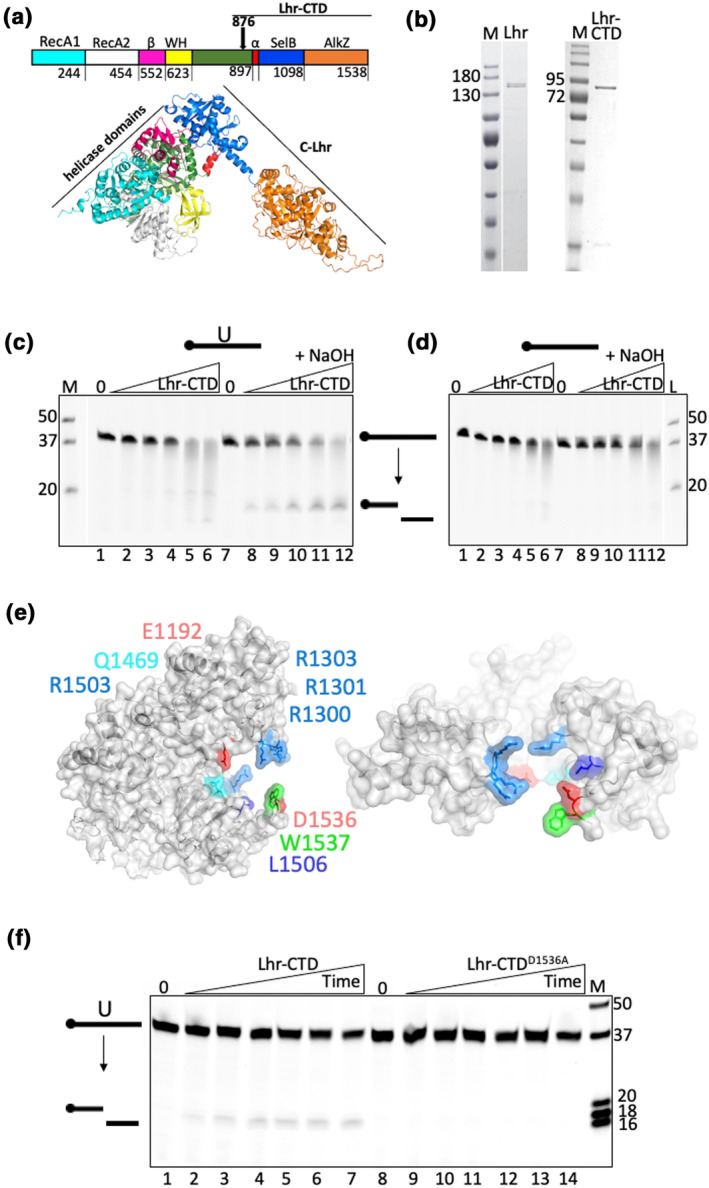
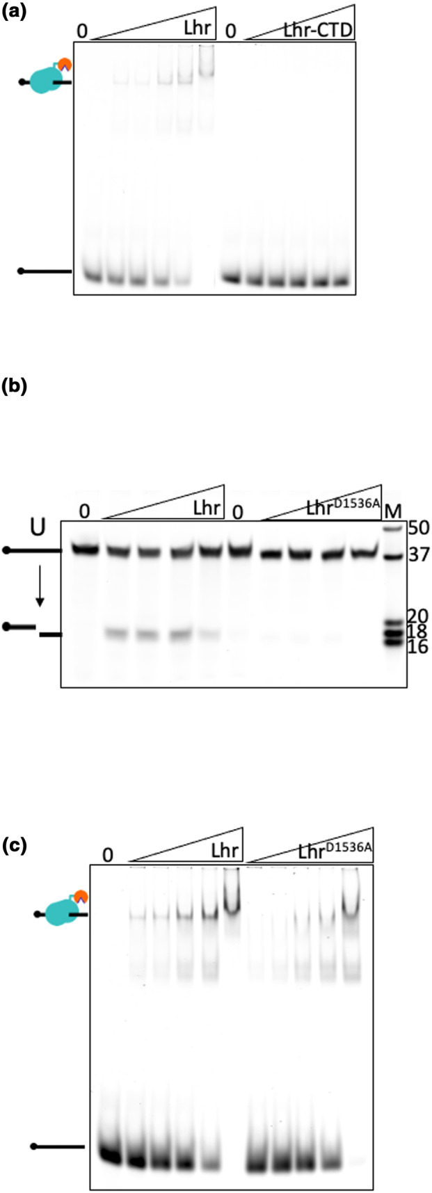
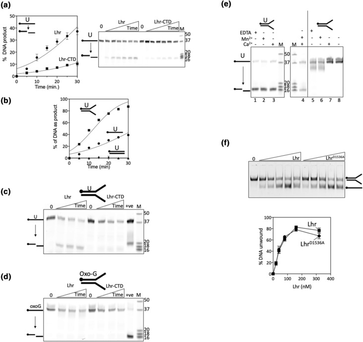
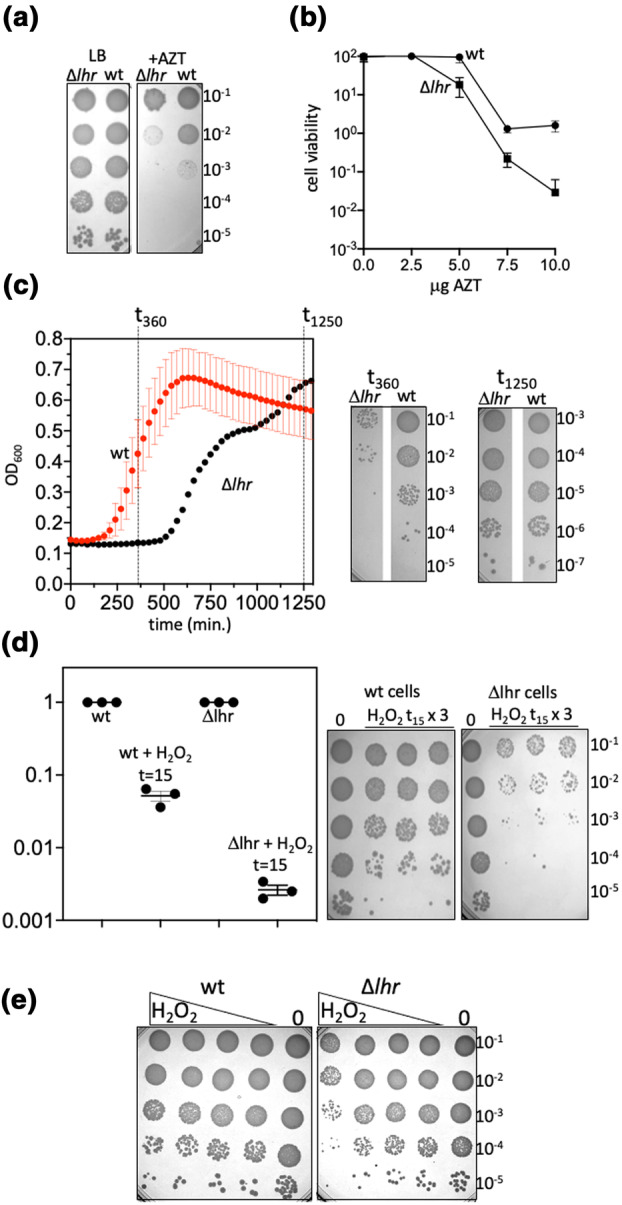
DNA glycosylases protect genetic fidelity during DNA replication by removing potentially mutagenic chemically damaged DNA bases. Bacterial Lhr proteins are well-characterized DNA repair helicases that are fused to additional 600-700 amino acids of unknown function, but with structural homology to SecB chaperones and AlkZ DNA glycosylases. Here, we identify that Escherichia coli Lhr is a uracil-DNA glycosylase (UDG) that depends on an active site aspartic acid residue. We show that the Lhr DNA helicase activity is functionally independent of the UDG activity, but that the helicase domains are required for fully active UDG activity. Consistent with UDG activity, deletion of lhr from the E. coli chromosome sensitized cells to oxidative stress that triggers cytosine deamination to uracil. The ability of Lhr to translocate single-stranded DNA and remove uracil bases suggests a surveillance role to seek and remove potentially mutagenic base changes during replication stress.

Keywords: DNA repair; DNA replication; glycosylase; helicase; uracil.


Figures

FIGURE 1
FIGURE 1
The E. coli Lhr‐CTD is a uracil‐DNA glycosylase requiring a catalytic aspartic acid. (a) AlphaFold 2 structural model of E. coli Lhr that is based on strong homology with the cryo‐EM structure of Lhr helicase core and Lhr‐CTD from M. smegmatis, respectively, PDB: 5V9X and PDB:7LHL. The E. coli Lhr‐Core helicase (amino acids 1–897) contains RecA domains, a beta‐sheet bundle (β), and a winged helix domain (WH) as indicated. Lhr‐CTD (amino acids 898–1538) comprises folds with structural homology to SecB chaperones and AlkZ glycosylases, as indicated. (b) Coomassie‐stained SDS‐PAGE acrylamide gels showing purified Lhr and Lhr‐CTD, with molecular mass ladder (M) values in kDa. (c) Products from mixing Lhr‐CTD (50, 100, 200, 400, and 800 nM) with 5′ Cy5‐ssDNA (12.5 nM) containing a d‐uracil base located 18 nucleotides from the fluorescent moiety as indicated (lanes 1–12), seen in a 15% denaturing acrylamide TBE gel. Addition of NaOH (lanes 8–12) causes β/δ elimination at the site of the abasic DNA product, resulting in DNA backbone cleavage. This confirms glycosylase protein activity. Marker (M) is made from known lengths of 5′ Cy5 ssDNA. (d) As for (c) in reactions containing unmodified 5′ Cy5‐ssDNA (12.5 nM). (e) Phyre2 structural model of E. coli Lhr‐CTD with predicted active site residues as labeled, including Lhr‐CTD residue Asp‐1536 mutated in this work. (f) Products from mixing Lhr‐CTD or Lhr‐CTDD1536A proteins (50 nM) with 12.5 mM d‐uracil containing 5′ Cy5‐ssDNA substrate, viewed in a 18% acrylamide denaturing TBE gel. Product formation is shown every 5 min for 30 min, observing no glycosylase activity from Lhr‐CTDD1536A.
FIGURE 2
FIGURE 2
LhrD1536A is inactive as a glycosylase but binds to DNA. (a) EMSA assays showing Lhr (12.5, 25, 50, 100, and 200 nM) complexes bound to single‐stranded DNA (12.5 nM, Supporting Information Table S2) that are stable migrating through a 5% acrylamide TBE gel (indicated to left of the gel as protein bound to ssDNA), compared with Lhr‐CTD at the same concentrations. (b) Products of Lhr glycosylase activity seen in an 18% acrylamide denaturing TBE gel were absent when reactions contained LhrD1536A. Proteins were used at 25, 50, 100, and 200 nM, with 12.5 nM of d‐uracil containing 5′ Cy5‐ssDNA substrate. (c) EMSA showing that LhrD1536A and Lhr (12.5, 25, 50, 100, and 200 nM) form stable complex with Cy5 end‐labeled single‐stranded DNA (Supporting Information Table S2) in a 5% acrylamide TBE gel.
FIGURE 3
FIGURE 3
Lhr is inactive against 8‐oxoguanine, and its uracil‐DNA glycosylase activity on duplex DNA functions independently from Lhr helicase activity. (a) Time‐dependent uracil‐DNA glycosylase activity of Lhr (50 nM) compared with Lhr‐CTD. The data show means of glycosylase activity (n = 3, with bars for standard error) alongside a representative gel that was used for quantification. The cartoon to the top left of the graph indicates Lhr‐catalyzed hydrolysis of the uracil‐containing ssDNA, as shown in gels in Figure 1. (b) Comparison of Lhr (50 nM) glycosylase activity on ss‐, ds‐, and forked d‐uracil containing DNA substrates (12.5 nM) as a function of time, with samples taken at time points indicated—plots are means of two independent experiments showing standard error bars. (c) Time‐course assays (10, 20, and 30 min) showing products from Lhr and Lhr‐CTD (each 80 nM) mixed with the preferred flayed duplex uracil‐DNA, seen in an 18% acrylamide denaturing TBE gel. Known length DNA strands are shown (M) and the positive control reaction (+ve) is product from 5 units of E. coli uracil‐DNA glycosylase. (d) As for (c) except d‐uracil‐DNA was replaced with otherwise identical 8‐oxo‐d‐Guanine DNA, and the control reaction (+ve) shows product formed by 5 units of formamidopyrimidine DNA glycosylase (Fpg) protein. (e) Lhr (80 nM) uracil‐DNA glycosylase activity seen as products in 18% acrylamide denaturing TBE gels (lanes 1–4), after 30‐minute reactions in either EDTA, manganese or calcium, each replacing magnesium as indicated, compared with unmodified DNA (lanes 5–8). (f) Shows a representative gel and graphical plot of DNA unwinding by Lhr and LhrD1536A proteins (20, 40, 80, 160, and 320 nM) on 12.5 nM of 5′ Cy5 labeled flayed duplex DNA, assessed in 10% acrylamide TBE gels. The data plot shows means of three experiments for each protein, showing standard error bars.
FIGURE 4
FIGURE 4
E. coli cells lacking Lhr are sensitive to oxidative stress. (a) Viability spot tests showing moderately increased sensitivity of Δlhr cells to AZT (7.5 μg/mL) compared with wild‐type (wt) cells, and; (b) represented in viability curves when Δlhr and wild‐type cells were grown independently in media containing AZT at 2.5, 5, 7.5, or 10 μg/mL. The plots show grow relative to wild‐type cells grown in media lacking AZT, as means of three experiments. (c) Growth of Δlhr and wild‐type cells monitored in 96‐well plates by optical density in media containing 5.8 mM H2O2, and with corresponding representative viability spot tests taken at the time points indicted during growth. (d) Viability of Δlhr and wild‐type cells in response to 15 min (t15) exposure to hydrogen peroxide. Growths were in triplicate, with a mean of three colony counts from wild‐type and Δlhr cells that were untreated, in the graph represented as 3 data points all at 1.0 as mean, and on the agar plates represented in the lane marked 0. Data points for colony counts after t15 hydrogen peroxide were calculated from the mean (1.0) untreated cultures and are shown as the three actual data values with standard error bars. The agar plates highlight the difference between wild‐type and Δlhr cells. (e) Viability spot tests comparing Δlhr and wild‐type cells grown without H2O2 to optical density prior to plating on to LB media containing 1.5625, 3.75, 6.25, and 12.5 mM H2O2.

Similar articles

References

    1. Almatarneh, M.H. , Flinn, C.G. & Poirier, R.A. (2008) Mechanisms for the deamination reaction of cytosine with H2O/OH(−) and 2H2O/OH(−): a computational study. Journal of Chemical Information and Modeling, 48(4), 831–843. - PubMed
    1. Aravind, L. & Koonin, E.V. (2000) The alpha/beta fold uracil DNA glycosylases: a common origin with diverse fates. Genome Biology, 1(4), RESEARCH0007. - PMC - PubMed
    1. Buckley, R.J. , Kramm, K. , Cooper, C.D.O. , Grohmann, D. & Bolt, E.L. (2020) Mechanistic insights into Lhr helicase function in DNA repair. The Biochemical Journal, 477(16), 2935–2947. - PMC - PubMed
    1. Buttner, K. , Nehring, S. & Hopfner, K.P. (2007) Structural basis for DNA duplex separation by a superfamily‐2 helicase. Nature Structural & Molecular Biology, 14(7), 647–652. - PubMed
    1. Cadet, J. & Wagner, J.R. (2013) DNA base damage by reactive oxygen species, oxidizing agents, and UV radiation. Cold Spring Harb Perspectives in Biology, 5(2). 10.1101/cshperspect.a012559 - DOI - PMC - PubMed

Publication types