. 2023 Jul 14;14(1):4196.
doi: 10.1038/s41467-023-39653-5.

Structural insights into the mechanism of overcoming Erm-mediated resistance by macrolides acting together with hygromycin-A

Affiliations

Structural insights into the mechanism of overcoming Erm-mediated resistance by macrolides acting together with hygromycin-A

Chih-Wei Chen et al. Nat Commun. .

Abstract

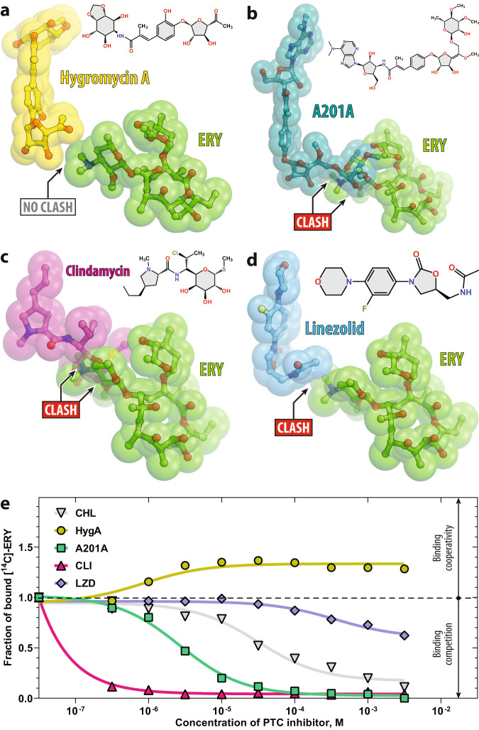
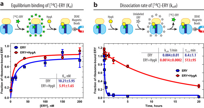
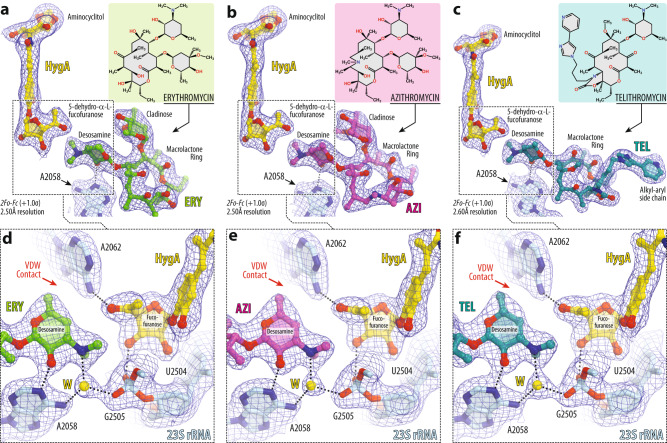
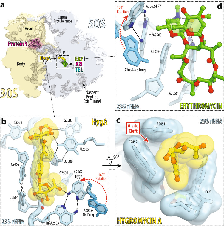
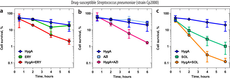
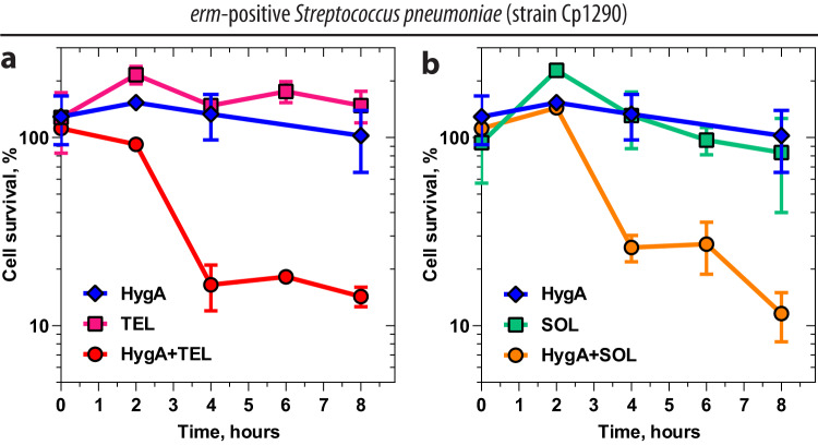
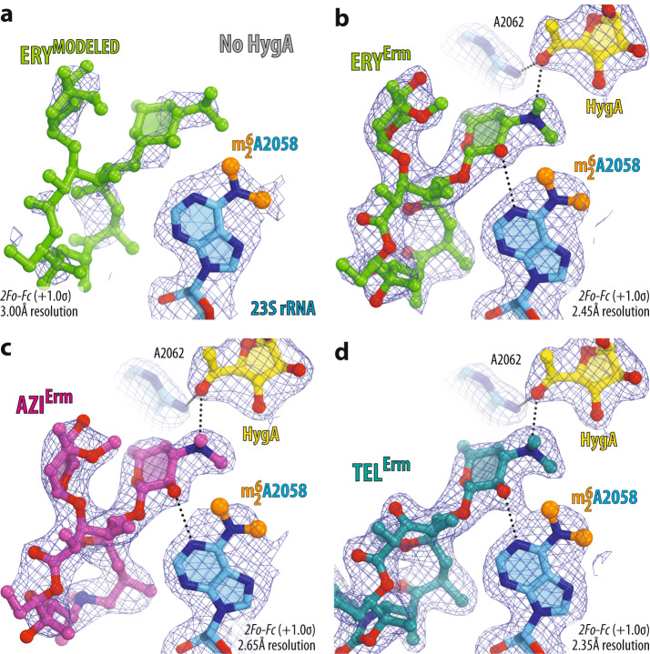
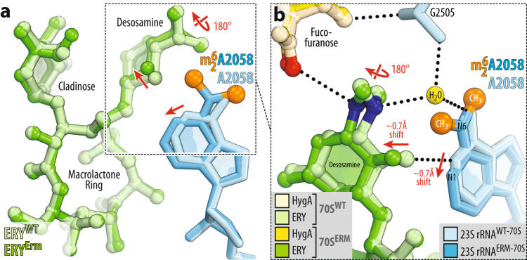
The ever-growing rise of antibiotic resistance among bacterial pathogens is one of the top healthcare threats today. Although combination antibiotic therapies represent a potential approach to more efficiently combat infections caused by susceptible and drug-resistant bacteria, only a few known drug pairs exhibit synergy/cooperativity in killing bacteria. Here, we discover that well-known ribosomal antibiotics, hygromycin A (HygA) and macrolides, which target peptidyl transferase center and peptide exit tunnel, respectively, can act cooperatively against susceptible and drug-resistant bacteria. Remarkably, HygA slows down macrolide dissociation from the ribosome by 60-fold and enhances the otherwise weak antimicrobial activity of the newest-generation macrolide drugs known as ketolides against macrolide-resistant bacteria. By determining a set of high-resolution X-ray crystal structures of drug-sensitive wild-type and macrolide-resistant Erm-methylated 70S ribosomes in complex with three HygA-macrolide pairs, we provide a structural rationale for the binding cooperativity of these drugs and also uncover the molecular mechanism of overcoming Erm-type resistance by macrolides acting together with hygromycin A. Altogether our structural, biochemical, and microbiological findings lay the foundation for the subsequent development of synergistic antibiotic tandems with improved bactericidal properties against drug-resistant pathogens, including those expressing erm genes.


Conflict of interest statement

The authors declare no competing interests.

Figures

Fig. 1
Fig. 1. Comparison of the macrolide binding site with those of various PTC-targeting antibiotics.
Superposition of the structures of the ribosome-bound ERY (red, PDB entry 6XHX [10.2210/pdb6XHX/pdb]) and a hygromycin A (yellow, PDB entry 5DOY [10.2210/pdb5DOY/pdb]), b nucleoside antibiotic A201A (teal, PDB entry 4Z3S [10.2210/pdb4Z3S/pdb]), c clindamycin (magenta, PDB entry 4V7V [10.2210/pdb4V7V/pdb]), or d linezolid (light blue, PDB entry 7S1G [10.2210/pdb7S1G/pdb]). All structures of ribosome-bound antibiotics were aligned based on domain V of the 23S rRNA. Note that only hygromycin A does not sterically clash with ribosome-bound ERY, while A201A, clindamycin, and linezolid overlap with the desosamine sugar of ERY. e Competition binding assay to assess the release of [14C]-radiolabeled ERY from the 70S ribosomes in the presence of increasing concentrations of one of the PTC-targeting antibiotics shown in (a)–(d). Chloramphenicol (CHL, gray) is used as a positive control known to compete with ERY. The amount of [14C]-ERY associated with the ribosomes in the absence of a competitor drug is arbitrarily assigned as 1.0 (dashed line). Under experimental conditions, this corresponds to ~50% of the 70S ribosomes bound to [14C]-ERY. The measurements were repeated twice with similar results. Source data are provided as a Source Data file. Note that, at high concentrations, only HygA (yellow) stimulates additional binding of ERY to the 70S ribosome, whereas A201A (teal), clindamycin (magenta), linezolid (light blue), or chloramphenicol (gray) cause its dissociation.
Fig. 2
Fig. 2. Effects of hygromycin A on binding properties of erythromycin to the 70S ribosome.
a Equilibrium binding of radiolabeled [14C]-ERY to determine dissociation constants (Kd). S. pneumoniae 70S ribosomes pre-equilibrated with increasing concentrations of [14C]-ERY in the absence (blue curve) and presence (red curve) of 100 μM HygA were isolated using positively-charged DEAE magnetic beads, and the amount of remaining ribosome-associated radioactivity was measured in a scintillation counter. Error bars show mean standard deviation of three independent measurements. Note that HygA does not significantly improve the binding affinity of ERY to the ribosome. b Kinetics of [14C]-ERY dissociation from S. pneumoniae 70S ribosomes saturated with [14C]-ERY in the absence (blue curve) and presence (red curve) of 100 μM HygA. The amount of remaining ribosome-associated radioactivity was measured at different time points after diluting ribosomes with molar excess of non-radiolabeled ERY. Experimental data were fitted with a one-phase exponential function that yielded dissociation rate constants (koff) of 0.084 ± 0.01 1/min for ERY alone and 0.0014 ± 0.0002 1/min for ERY in the presence of HygA. The constants were used to calculate the half-lives (t1/2) of the complexes (shown in the figure). Error bars show mean standard deviation of two independent measurements. Source data are provided as a Source Data file. Note that HygA significantly (60-fold) slows down the dissociation rate of ERY from the 70S ribosome.
Fig. 3
Fig. 3. Electron density maps of three macrolides bound to the T. thermophilus 70S ribosome together with hygromycin A.
2Fo-Fc electron difference Fourier maps of hygromycin A (HygA, yellow) and either erythromycin (a, ERY, green), or azithromycin (b, AZI, magenta), or telithromycin (c, TEL, teal). The refined models of antibiotics are displayed in their respective electron density maps after the refinement (blue mesh). The overall resolution of the corresponding structures and the contour levels of the depicted electron density maps are shown in the bottom left corner of each panel. Chemical structures of corresponding macrolides are shown as insets. Close-up views of high-resolution electron density maps of ribosome-bound HygA together with ERY (d), AZI (e), or TEL (f) interacting with the nucleotides of the 23S rRNA (light blue). Note that the dimethylamino group of all macrolides is rotated toward nucleotide A2058 and forms an H-bond with a water molecule (W, yellow) tightly coordinated by the exocyclic N6-amino group of A2058 and the phosphate of G2505. Nitrogens are colored blue; oxygens are red (except for the water). Also, note that the atoms of desosamine group of a macrolide form van der Waals contacts with the fucofuranose moiety of HygA and nucleobase A2062 (red arrows).
Fig. 4
Fig. 4. Structure of HygA and ERY simultaneously bound to the 70S ribosome.
a Overview of the T. thermophilus 70S ribosome structure with bound HygA (yellow) and ERY (green) viewed as a cross-cut section through the nascent peptide exit tunnel (NPET). The 30S subunit is shown in light yellow; the 50S subunit is in light blue; ribosome-bound protein Y is colored magenta. The binding positions of AZI and TEL are nearly the same as ERY. Close-up views of the HygA (b, c) and ERY (d) binding sites in the PTC and NPET of the 70S ribosome, respectively (E. coli numbering of the rRNA nucleotides is used throughout). H-bond interactions are indicated with dotted lines. Note that by forming an H-bond with the base of nucleotide A2062 of the 23S rRNA (light blue), HygA causes rotation of this nucleotide by ~160 degrees to form Hoogsteen base-pair with the m2A2503 residue of the 23S rRNA (red dashed arrow). The binding of ERY, as well as other macrolides, causes the same characteristic rotation of nucleotide A2062. The unrotated conformation of A2062 observed in the absence of either drug is shown in blue (PDB entry 4Y4O [10.2210/pdb4Y4O/pdb]).
Fig. 5
Fig. 5. Hygromycin A potentiates bactericidal properties of macrolide antibiotics.
Time-kill assays using drug-susceptible Cp2000 strain of S. pneumoniae exposed during various times to antibiotic concentrations at 4x MIC of hygromycin A (HygA, blue plot, 20 µg/ml), macrolides erythromycin (a, ERY, green plot, 0.25 µg/ml), azithromycin (b, AZI, light blue plot, 0.5 µg/ml), or ketolide solithromycin (c, SOL, teal plot, 0.04 µg/ml) alone and in combination with HygA (red, magenta, and orange plots, respectively). The initial number of viable cells (colony-forming units, CFUs) before the addition of drug(s) was arbitrarily assigned to 100%. Error bars show mean standard deviation of 2–4 independent measurements. Source data are provided as a Source Data file.
Fig. 6
Fig. 6. Hygromycin A restores the bactericidal activity of ketolides against Erm-expressing bacteria.
Time-kill assays using erm-positive macrolide-resistant Cp1290 strain of S. pneumoniae exposed during various times to antibiotic concentrations at 4x MIC of hygromycin A (HygA, blue plot, 20 µg/ml), ketolides telithromycin (a, TEL, magenta plot, 2 µg/ml) or solithromycin (b, SOL, teal plot, 4 µg/ml) alone and in combination with HygA (red and orange plots, respectively). The initial number of viable cells (colony-forming units, CFUs) before the addition of drug(s) was arbitrarily assigned to 100%. Error bars show mean standard deviation of 2–5 independent measurements. Source data are provided as a Source Data file.
Fig. 7
Fig. 7. Electron density maps of three macrolides bound to the Erm-modified 70S ribosome in the presence of hygromycin A.
Electron density maps of macrolides erythromycin (a, b), azithromycin (c), or telithromycin (d) in the absence (a) and presence of HygA (bd) bound to the Erm-modified Tth 70S ribosome containing N6-dimethylated residue A2058 (blue with methyl groups highlighted in orange) in the 23S rRNA. H-bonds are depicted with dotted lines.
Fig. 8
Fig. 8. Comparison of structures of erythromycin bound to the WT and Erm-modified 70S ribosome in the presence of hygromycin A.
a, b Superposition of ERY (light green) in complex with the WT 70S ribosome containing unmodified residue A2058 (light blue) and the structure of ERY (green) in complex with the Erm-modified 70S ribosome containing N6-dimethylated residue A2058 (blue with methyl groups highlighted in orange). Note that, while the overall position of ribosome-bound ERY is nearly identical in the two structures, N6-dimethylation of A2058 results in a 180-degree rotation of the dimethylamino group of desosamine away from A2058 nucleobase toward the fucofuranose moiety of HygA and formation of a direct H-bond with it. Also, note that N6-dimethylation of A2058 causes a small shift of ERY’s desosamine moiety away from the nucleotide, potentially weakening the H-bond between the 2’-OH of macrolide and the N1 atom of A2058 (red arrows).

References

    1. Provenzani A, et al. Multidrug-resistant gram-negative organisms: a review of recently approved antibiotics and novel pipeline agents. Int. J. Clin. Pharm. 2020;42:1016–1025. doi: 10.1007/s11096-020-01089-y. - DOI - PubMed
    1. Bernal FA, Hammann P, Kloss F. Natural products in antibiotic development: is the success story over? Curr. Opin. Biotechnol. 2022;78:102783. doi: 10.1016/j.copbio.2022.102783. - DOI - PubMed
    1. Konwar AN, Hazarika SN, Bharadwaj P, Thakur D. Emerging non-traditional approaches to combat antibiotic resistance. Curr. Microbiol. 2022;79:330. doi: 10.1007/s00284-022-03029-7. - DOI - PMC - PubMed
    1. Lewis K. The science of antibiotic discovery. Cell. 2020;181:29–45. doi: 10.1016/j.cell.2020.02.056. - DOI - PubMed
    1. Imai Y, et al. A new antibiotic selectively kills Gram-negative pathogens. Nature. 2019;576:459–464. doi: 10.1038/s41586-019-1791-1. - DOI - PMC - PubMed

Publication types

MeSH terms