. 2023 Jul 14;13(1):11423.
doi: 10.1038/s41598-023-38733-2.

Surface characterization, electrochemical properties and in vitro biological properties of Zn-deposited TiO2 nanotube surfaces

Affiliations

Surface characterization, electrochemical properties and in vitro biological properties of Zn-deposited TiO2 nanotube surfaces

Salih Durdu et al. Sci Rep. .

Abstract

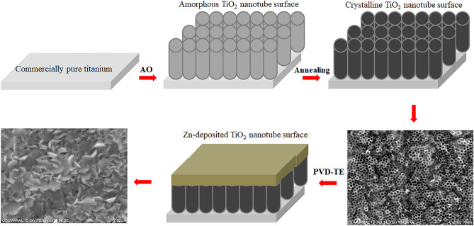
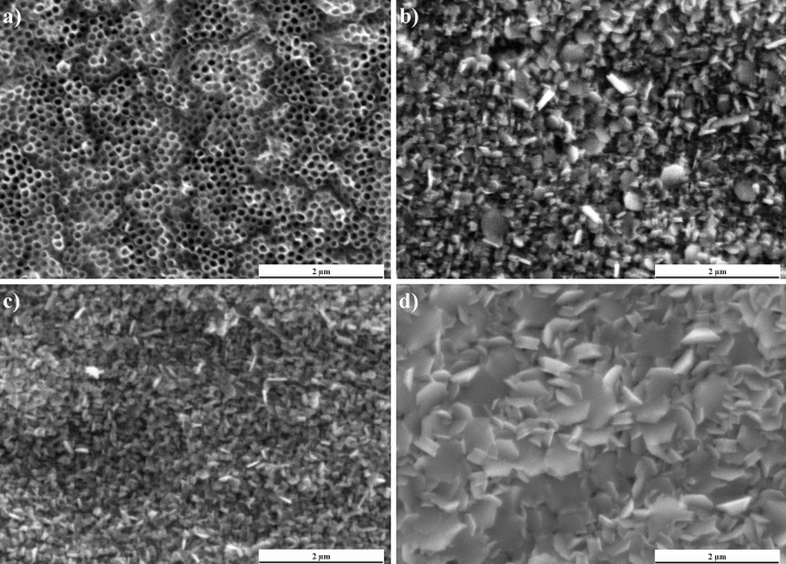
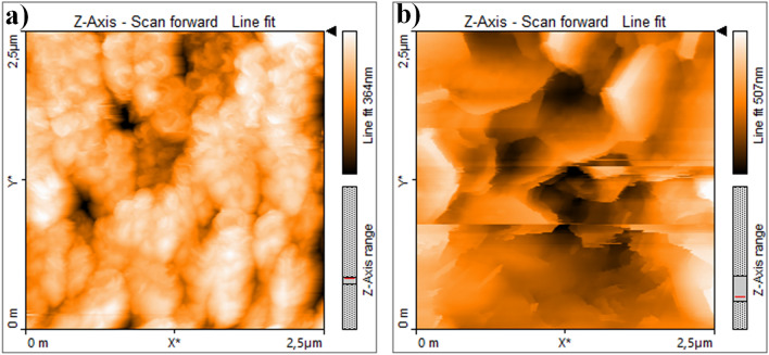
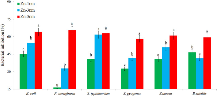
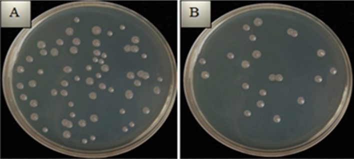
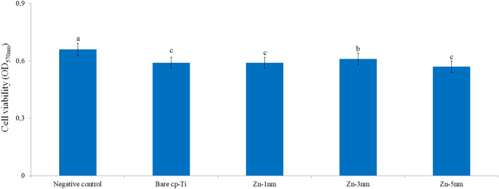
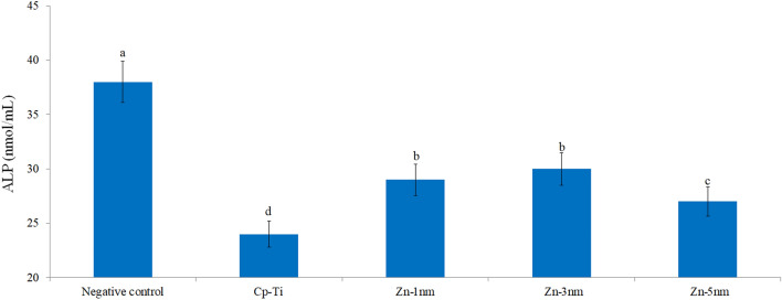
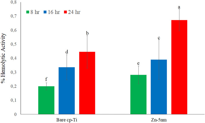
In this work, to improve antibacterial, biocompatible and bioactive properties of commercial pure titanium (cp-Ti) for implant applications, the Zn-deposited nanotube surfaces were fabricated on cp-Ti by using combined anodic oxidation (AO) and physical vapor deposition (PVD-TE) methods. Homogenous elemental distributions were observed through all surfaces. Moreover, Zn-deposited surfaces exhibited hydrophobic character while bare Ti surfaces were hydrophilic. Due to the biodegradable behavior of Zn on the nanotube surface, Zn-deposited nanotube surfaces showed higher corrosion current density than bare cp-Ti surface in SBF conditions as expected. In vitro biological properties such as cell viability, ALP activity, protein adsorption, hemolytic activity and antibacterial activity for Gram-positive and Gram-negative bacteria of all surfaces were investigated in detail. Cell viability, ALP activity and antibacterial properties of Zn-deposited nanotube surfaces were significantly improved with respect to bare cp-Ti. Moreover, hemolytic activity and protein adsorption of Zn-deposited nanotube surfaces were decreased. According to these results; a bioactive, biocompatible and antibacterial Zn-deposited nanotube surfaces produced on cp-Ti by using combined AO and PVD techniques can have potential for orthopedic and dental implant applications.


Conflict of interest statement

The authors declare no competing interests.

Figures

Figure 1
Figure 1
Schematic representation of fabrication of ZnO-based TiO2 nanotube arrays on cp-Ti by AO and PVD-TE processes.
Figure 2
Figure 2
Surface SEM images: (a) bare nanotube surface, (b) Zn-1 nm, (c) Zn-3 nm and (d) Zn-5 nm.
Figure 3
Figure 3
EDX-mapping images of Zn-deposited nanotube surfaces: (a) Zn-1 nm, (b) Zn-3 nm and (c) Zn-5 nm.
Figure 4
Figure 4
XRD spectra of bare nanotube and Zn-deposited nanotube surfaces: (a) bare nanotube, (b) Zn-1 nm, (c) Zn-3 nm and (d) Zn-5 nm.
Figure 5
Figure 5
AFM images: (a) bare nanotube and (b) Zn-deposited nanotube surfaces.
Figure 6
Figure 6
Contact angle measurement images: (a) bare cp-Ti, (b) bare nanotube, (c) Zn-1 nm, (d) Zn-3 nm and (e) Zn-5 nm.
Figure 7
Figure 7
Percentage of microbial inhibition of Zn-deposited nanotube surfaces for Gram-positive and Gram-negative bacteria. Values shown with different letters for each bacteria are statistically significant.
Figure 8
Figure 8
Decrease in bacterial colonies after recultivation in samples with the highest antibacterial activity on Zn-deposited surfaces: (a) P. aeruginosa viability after reculturation on bare cp-Ti surfaces, (b) P. aeruginosa viability after reculture on Zn-5 nm surfaces.
Figure 9
Figure 9
MTT test results of the bare cp-Ti and Zn-deposited nanotube surfaces after 4 h of incubation. Values shown with different letters are statistically significant.
Figure 10
Figure 10
ALP activity in cells adhering to bare cp-Ti and Zn-deposited nanotube surfaces. Values shown with different letters are statistically significant.
Figure 11
Figure 11
Hemolytic activities of bare cp-Ti and Zn-deposited (Zn-5 nm) nanotube surfaces for 8 h, 16 h and 24 h. Values shown with different letters are statistically significant.

References

    1. Rios J, et al. Self-organized TiO2 nanotubes on Ti–Nb–Fe alloys for biomedical applications: Synthesis and characterization. Electrochem. Commun. 2022;138:107280. doi: 10.1016/j.elecom.2022.107280. - DOI
    1. Kulkarni M, et al. Titanium nanostructures for biomedical applications. Nanotechnology. 2015 doi: 10.1088/0957-4484/26/6/062002. - DOI - PubMed
    1. Niinomi M, Nakai M, Hieda J. Development of new metallic alloys for biomedical applications. Acta Biomater. 2012;8:3888–3903. doi: 10.1016/j.actbio.2012.06.037. - DOI - PubMed
    1. Jaafar A, Hecker C, Arki P, Joseph Y. Sol–gel derived hydroxyapatite coatings for titanium implants: A review. Bioeng. Basel. 2020 doi: 10.3390/bioengineering7040127. - DOI - PMC - PubMed
    1. Baltatu MS, Sandu AV, Nabialek M, Vizureanu P, Ciobanu G. Biomimetic deposition of hydroxyapatite layer on titanium alloys. Micromachines. 2021;12:1447. doi: 10.3390/mi12121447. - DOI - PMC - PubMed

Publication types

MeSH terms