. 2023 Jul 14;118(1):28.
doi: 10.1007/s00395-023-00997-0.

Endothelial ILK induces cardioprotection by preventing coronary microvascular dysfunction and endothelial-to-mesenchymal transition

Affiliations

Endothelial ILK induces cardioprotection by preventing coronary microvascular dysfunction and endothelial-to-mesenchymal transition

P Reventun et al. Basic Res Cardiol. .

Abstract

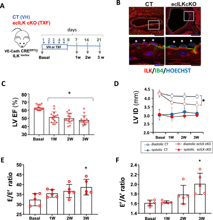
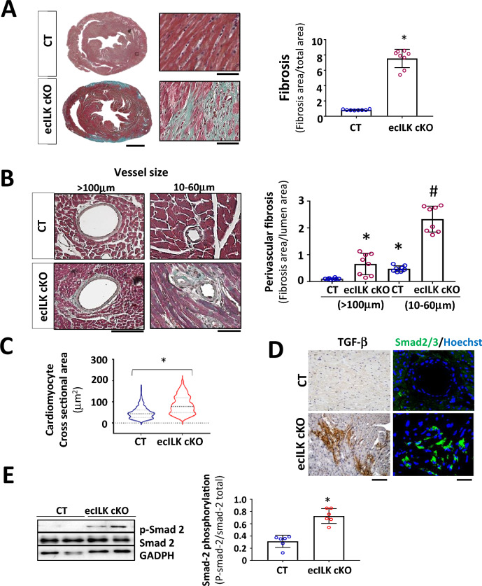
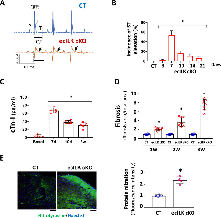
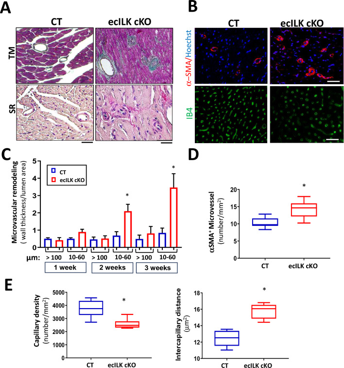
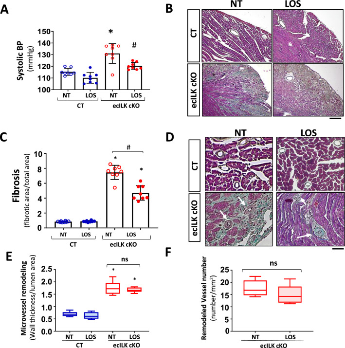
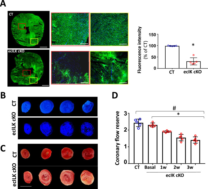
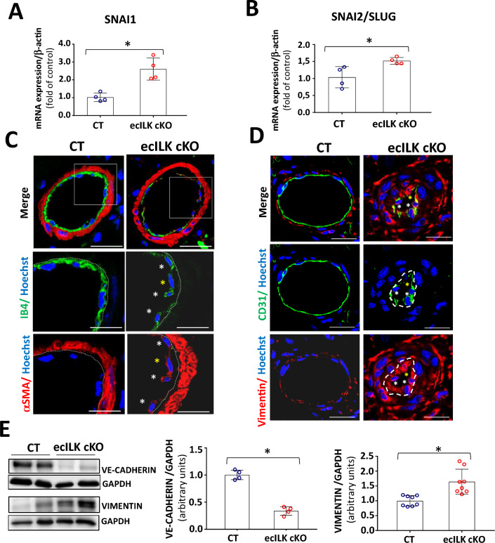
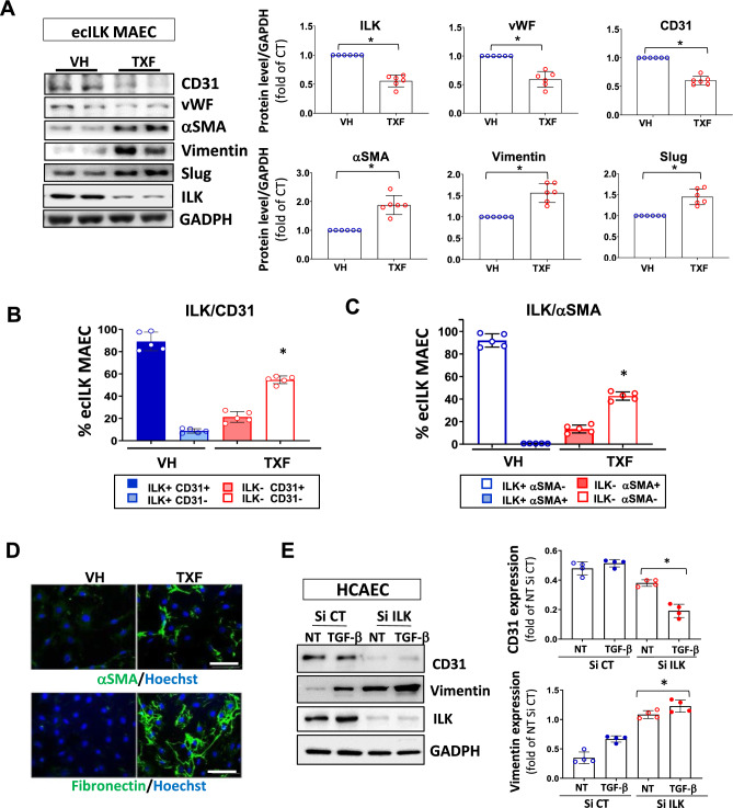
Endothelial dysfunction is an early event in coronary microvascular disease. Integrin-linked kinase (ILK) prevents endothelial nitric oxide synthase (eNOS) uncoupling and, thus, endothelial dysfunction. However, the specific role of endothelial ILK in cardiac function remains to be fully elucidated. We hypothesised that endothelial ILK plays a crucial role in maintaining coronary microvascular function and contractile performance in the heart. We generated an endothelial cell-specific ILK conditional knock-out mouse (ecILK cKO) and investigated cardiovascular function. Coronary endothelial ILK deletion significantly impaired cardiac function: ejection fraction, fractional shortening and cardiac output decreased, whilst left ventricle diastolic internal diameter decreased and E/A and E/E' ratios increased, indicating not only systolic but also diastolic dysfunction. The functional data correlated with extensive extracellular matrix remodelling and perivascular fibrosis, indicative of adverse cardiac remodelling. Mice with endothelial ILK deletion suffered early ischaemic-like events with ST elevation and transient increases in cardiac troponins, which correlated with fibrotic remodelling. In addition, ecILK cKO mice exhibited many features of coronary microvascular disease: reduced cardiac perfusion, impaired coronary flow reserve and arterial remodelling with patent epicardial coronary arteries. Moreover, endothelial ILK deletion induced a moderate increase in blood pressure, but the antihypertensive drug Losartan did not affect microvascular remodelling whilst only partially ameliorated fibrotic remodelling. The plasma miRNA profile reveals endothelial-to-mesenchymal transition (endMT) as an upregulated pathway in endothelial ILK conditional KO mice. Our results show that endothelial cells in the microvasculature in endothelial ILK conditional KO mice underwent endMT. Moreover, endothelial cells isolated from these mice and ILK-silenced human microvascular endothelial cells underwent endMT, indicating that decreased endothelial ILK contributes directly to this endothelial phenotype shift. Our results identify ILK as a crucial regulator of microvascular endothelial homeostasis. Endothelial ILK prevents microvascular dysfunction and cardiac remodelling, contributing to the maintenance of the endothelial cell phenotype.

Keywords: Endothelial to mesenchymal transition; Fibrosis; Integrin-linked kinase; Ischemia; Microvascular dysfunction; Microvessel remodeling.


Conflict of interest statement

The authors declare that they have no conflict of interest.

Figures

Fig. 1
Fig. 1
ILK deletion in coronary endothelium leads to myocardial dysfunction. A Schematic figure showing the experimental design. VE-Cadh-CreERT2/ILKlox/lox mice were treated (i.p.) during 5 days with Tamoxifen;TXF resulting in endothelial ILK deletion (ecILK cKO in red) or vehicle;VH (CT in blue). Cardiovascular function was tested before treatment (basal) and once a week up to three weeks. Hearts were harvested at 1, 2 and 3 weeks. B Representative confocal images of a coronary artery in CT (left panel) and ecILK cKO mice at three weeks stained with anti-ILK (red), endothelial cells were stained with IB4 (green) and nuclei were counterstained in blue with Hoechst. Asterisks in magnification indicate individual endothelial cells showing no expression of ILK (red) in the tamoxifen treated group. Scale bar (upper panel): 25 mm and 10 μm lower panel. C Bar graph showing the ejection fraction of the left ventricle (LV EF) in ecILK cKO mice (n = 15, *p < 0.001 vs Basal). D Changes in the internal diameter of left ventricle in diastole (LVIDd) and systole (LVIDs) in CT and ecILK cKO mice. (n = 15, *p < 0.005 vs CT diastole at three weeks). E. E/Eʹ ratio (LV end–diastolic filling pressure) and F Eʹ/Aʹ ratio from ecILKcKO mice (n = 5, *p < 0.001 vs Basal). All p values were calculated using ANOVA
Fig. 2
Fig. 2
Endothelial-specific ILK deletion leads to adverse remodelling. A Representative Masson Trichrome staining of CT and ecILK cKO mice heart sections (Scale bar = 1 mm) showing cardiac lesions three weeks after deletion. Right: Magnification of ecILK cKO and CT hearts. Scale bar: 50 μm. Right panel: quantitative analysis of cardiac fibrosis. (n = 8,*p < 0, 005 vs CT). B Representative photo-micrographs from ecILK cKO and CT hearts stained with Masson Trichrome showing perivascular fibrosis in myocardial arteries > 100 μm diameter and small arterioles (60–10 μm). Scale bar: 100 μm (left panel); 50 μm (right panel). Right, quantitative analysis of perivascular fibrosis in vessels diameter > 100 μm and small arterioles (60–10 μm). (n = 8,*p < 0.001 vs CT > 100 μm; #p < 0, 005 vs CT small vessels). C Quantitative data of cardiomyocyte (CM) cell surface area; n = 6–10 hearts per group with 300–600 CMs analysed per heart. CM area is expressed in μm2. *p < 0, 05 CT vs ecILK cKO. D Representative heart sections of CT and ecILK cKO mice staining of TGF-β1 and SMAD2/3 (n = 8). Scale bar: 50 μm left and 25 μm right. E Immunoblot detection of phosphorylated Smad-2 in total heart lysates from CT and ecILK cKO mice. GAPDH was used as a loading control. Densitometric analysis (right panel) shows a significant increase in the expression of p-Smad2 in ecILK cKO mice (n = 6, *p < 0.05 vs CT). p values were calculated using Student’s t test and one-way ANOVA in B
Fig. 3
Fig. 3
ILK deletion in coronary endothelium promotes myocardial ischaemia. A Representative ECG recording (lead II) of CT (upper panel) and ecILK cKO mice (lower panel) 1 week after treatment, showing ST elevation (arrow) in ecILK cKO mice. B Incidence of ST elevation in CT (21d) and ecILK cKO mice at different time points post-deletion in six different experimental groups (5–8 mice/group;*p < 0,001 vs CT). C Plasmatic cardiac troponin quantitation showing a significant increase 7 days after treatment when compared to basal conditions and remain elevated up to three weeks after deletion (n = 6 mice per condition; *p < 0.001 vs Basal). D Myocardial Fibrosis from CT and ecILK cKO mice 1, 2 and 3 weeks after deletion/treatment (n = 8 mice per condition) *p < 0.05 vs CT at the same time point. E Representative heart sections from CT and ecILK cKO mice 3 weeks after deletion stained with nitrotyrosine (green), nuclei were counterstained with Hoetchst (scale bar: 50 μm). Right panel, quantitative analysis showed a significant increase in protein nitration expressed as relative fluorescence intensity in ecILK cKO mice (n = 5, *p < 0.05). p values were calculated using one-way ANOVA and Student’s t test in (E)
Fig. 4
Fig. 4
ILK endothelial deletion leads to altered microvascular structure and density. A Representative photomicrographs of heart sections stained with Masson Trichrome (upper panel) and Sirius Red (lower panel) showing the remodelled microvasculature in ecILK cKO (right panel) as compared to CT mice (left panel). Scale bar = 25 μm. B Representative photomicrographs of heart sections obtained from CT and ecILK cKO mice showing (upper panel) arterioles stained for smooth muscle actin (α-SMA, red) and counterstained in blue with Hoechst and (lower panel) capillaries stained for IB4 (green) Scale bar: 100 mm (n = 8). C Arteriolar wall thickness-to-lumen area ratio was significantly increased in ecILK cKO mice 2- and 3-weeks post-deletion only in small vessels (10–60 mm) and remained unchanged in large vessels (> 100 mm) (n = 8, *p < 0.05 vs CT). D Total number of α-SMA-positive microvessels (10-60 μm) was significantly increased in ecILK cKO mice three weeks after deletion (n = 6, *p < 0.05 vs CT). E In ecILK cKO mice, a significant decrease in capillary density (left panel) together with a significant increase in intercapillary distance (right panel) was observed 3 weeks after treatment when compared to CT mice (n = 6, *p < 0.05 vs CT). p values were calculated using Student’s t test and one-way ANOVA in (C)
Fig. 5
Fig. 5
Effect of the antihypertensive drug Losartan, on the myocardial and microvascular remodelling of ecILK cKO mice. CT and ecILK cKO mice were treated with Losartan (LOS, n = 8) or left untreated (NT, n = 8). A Three weeks after treatment, systolic arterial blood pressure was significantly increased in untreated ecILKcKO as compared to CT mice; this increase was prevented by LOS (*p < 0.05 vs CT; # p < 0.05 vs NT ecILK cKO). B Representative photomicrographs of heart sections obtained from CT and ecILK cKO mice treated as in A, stained with Masson Trichrome showing myocardial fibrosis at three weeks (n = 8) Scale bar: 100 μm. C Quantitative analysis of myocardial fibrosis at 3 weeks showing partial prevention in LOS treated vs NT treated ecILKcKO mice (n = 8, *p < 0.05 CT vs ecILK cKO; #p < 0.01 vs NT ecILK cKO). D Representative photomicrographs of heart sections obtained from CT and ecILK cKO mice treated as in A, stained with Masson Trichrome showing remodelled microvasculature (n = 8, scale bars: 25 μm, white arrows mark the remodelled vessels). E In ecILK cKO mice, the increased arteriolar wall thickness-to-lumen area ratio (in vessels between 10 and 60 mm of diameter) observed three weeks after deletion was unaffected by LOS (*p < 0.001 vs CT, n = 8). F The increased number of remodelled microvessels in 3 weeks ecILKcKO mice was unaffected by LOS (*p < 0.001 vs CT, n = 8). Differences amongst treatment groups were assessed by ANOVA and Student’s t test (F)
Fig. 6
Fig. 6
ILK deletion from coronary endothelium promoted microvascular dysfunction. A Representative confocal microscopy image of cardiac sections following in vivo dextran-fluorescein isothiocyanate (FITC) injection at 2 weeks after endothelial ILK deletion showing the absence of dextran-FITC (red box) and dextran-FITC accumulation (yellow box) in ecILK cKO microvessels compared to CT mice. Scale bar: 1 mm. Scale bar at magnifications: 100 mm. Right: fluorescence intensity quantitation expressed as percentage of CT (n = 6, * p < 0.005 vs CT). B and C Images show Thioflavin S and TTC staining in CT and ecILK cKO mice heart slices three weeks post deletion. B Under ultra-violet light, non-fluorescent regions on the Thioflavin S stain highlight areas of compromised coronary blood flow. (n = 4). C TTC sections indicate region of necrosis appearing white in the ecILK cKO group; CT group did no show infarction (n = 4). Scale bar = 5 mm. D Coronary flow reserve assessed by echocardiography at baseline and hyperemia in CT (3W) and ecILK cKO mice before (Basal) and at different time points after deletion showing decreased CFR at three weeks compared to CT and Basal (n = 5; *p < 0.001 vs Basal; #p < 0.001 vs CT). p values groups were assessed by ANOVA
Fig. 7
Fig. 7
Endothelial ILK deletion induces endMT in coronary microvasculature. RT-qPCR of CT and ecILK cKO mice cardiac extracts showing mRNA expression of endMT transcription factors A Snai1 and B Snai2 (Slug) are significantly increased in ecILKcKO mice (*p < 0.05, n = 4). C Representative confocal photomicrographs showing arterioles in cardiac sections from CT and ecILK cKO mice immunostained for IB4 (green) and α-SMA (red). Nuclei were counterstained with Hoechst (blue). White asterisks mark endothelial cells expressing both endothelial and mesenchymal cell markers. Yellow asterisk mark an endothelial cell expressing mostly α-SMA. (Scale bar: 25 mm, n = 6). D Representative confocal photomicrographs showing small arterioles from CT and ecILK cKO mice immunostained for CD31 (green) and Vimentin (red). Nuclei were counterstained with Hoechst (blue). (Scale bar: 10 μm, n = 6). E Western blot analysis of VE-cadherin and Vimentin protein expression in CT and ecILK cKO cardiac protein extracts. GAPDH was used as a loading control. Left panel shows representative blots. Quantitative analysis shows a significant decrease of both proteins in ecILK cKO mice (*p < 0.05, n = 4–8 mice). p values were calculated using Student’s t test
Fig. 8
Fig. 8
ILK deletion triggers endMT in ecILK MAEC and human coronary endothelial cells. A Western blot detection of endothelial (CD31 and vWF) and mesenchymal markers (αSMA and vimentin) expression, Slug and ILK in mouse aortic endothelial cells isolated from Cadh5-PAC-CRE ERT2/ILK flox mice (ecILK MAEC). ILK deletion was induced by treating the cells with Tamoxifen (10—6 M) (TXF) or vehicle (VH) for three days. GAPDH protein expression was used as loading control. In the right panel the quantitative analysis shows a significant decrease of ILK, vWF and CD31 in TXF ecILK MAEC as compared to VH (*p < 0.05, n = 6) and a significant increase in a-SMA, Vimentin and Slug (*p < 0.05, n = 6). Flow cytometry analysis of B endothelial marker CD31 and ILK and C ILK and α-SMA in VH and TXF treated ecILK MAEC. Figures represent the percentage of cells positive to each marker. VH-treated endothelial cells express CD31 and no α-SMA, whilst half of the TXF treated ecILK cells lose CD31 expression (ILK-,CD31-) whilst they become positive for α-SMA (ILK-, α-SMA +) (n = 5). *p < 0.01 vs CT. D Representative confocal microphotographs from VH and TXF ecILK MAEC stained for α-SMA (upper panel) or Fibronectin (lower panel). Nuclei were counterstained with Hoechst (blue). Scale bar: 10 μm. E Human coronary endothelial cells (HCAEC) were transfected with ILK-specific siRNA (si-ILK) or non-silencing siRNA scramble (si CT) and then, treated with 2 ng/ml TGFβ for 3 days. NT: non-treated. Left panel shows a representative immunoblot for the endothelial cell marker, CD31 and the mesenchymal cell marker, vimentin. GADPH protein expression is used as a loading control. Quantitative analysis (right panel) shows a significant reduction in CD31 expression when ILK was silenced which was further decreased after TGF-β treatment. Vimentin expression was increased in si-ILK cells, and further enhance after TGF- b treatment (*p < 0.05, n = 4) Blue circles: SiRNA CT/Red circles: SiRNA ILK. Empty: non-treated, filled: TGF-β treated.*p < 0.05 vs SiCT. p values were calculated using ANOVA

References

    1. Alvandi Z, Bischoff J. Endothelial-mesenchymal transition in cardiovascular disease. Arterioscler Thromb Vasc Biol. 2021;41:2357–2369. doi: 10.1161/ATVBAHA.121.313788. - DOI - PMC - PubMed
    1. Brutsaert DL. Cardiac endothelial-myocardial signaling: its role in cardiac growth, contractile performance, and rhythmicity. Physiol Rev. 2003;83:59–115. doi: 10.1152/PHYSREV.00017.2002. - DOI - PubMed
    1. Chevalier J, Yin H, Arpino JM, O’Neil C, Nong Z, Gilmore KJ, Lee JJ, Prescott E, Hewak M, Rice CL, Dubois L, Power AH, Hamilton DW, Pickering JG. Obstruction of small arterioles in patients with critical limb ischemia due to partial endothelial-to-mesenchymal transition. iScience. 2020 doi: 10.1016/J.ISCI.2020.101251. - DOI - PMC - PubMed
    1. Cuadrado I, Castejon B, Martin AM, Saura M, Reventun-Torralba P, Zamorano JL, Zaragoza C. Nitric oxide induces cardiac protection by preventing extracellular matrix degradation through the complex caveolin-3/emmprin in cardiac myocytes. PLoS ONE. 2016 doi: 10.1371/journal.pone.0162912. - DOI - PMC - PubMed
    1. Cuadrado I, Piedras MJGM, Herruzo I, del Carmen TM, Castejón B, Reventun P, Martin A, Saura M, Zamorano JL, Zaragoza C. EMMPRIN-targeted magnetic nanoparticles for in vivo visualization and regression of acute myocardial infarction. Theranostics. 2016;6:545–557. doi: 10.7150/THNO.13352. - DOI - PMC - PubMed

Publication types

Substances