. 2023 Jul 15;14(7):436.
doi: 10.1038/s41419-023-05964-0.

LRRK2 phosphorylation status and kinase activity regulate (macro)autophagy in a Rab8a/Rab10-dependent manner

Affiliations

LRRK2 phosphorylation status and kinase activity regulate (macro)autophagy in a Rab8a/Rab10-dependent manner

Elżbieta Kania et al. Cell Death Dis. .

Abstract

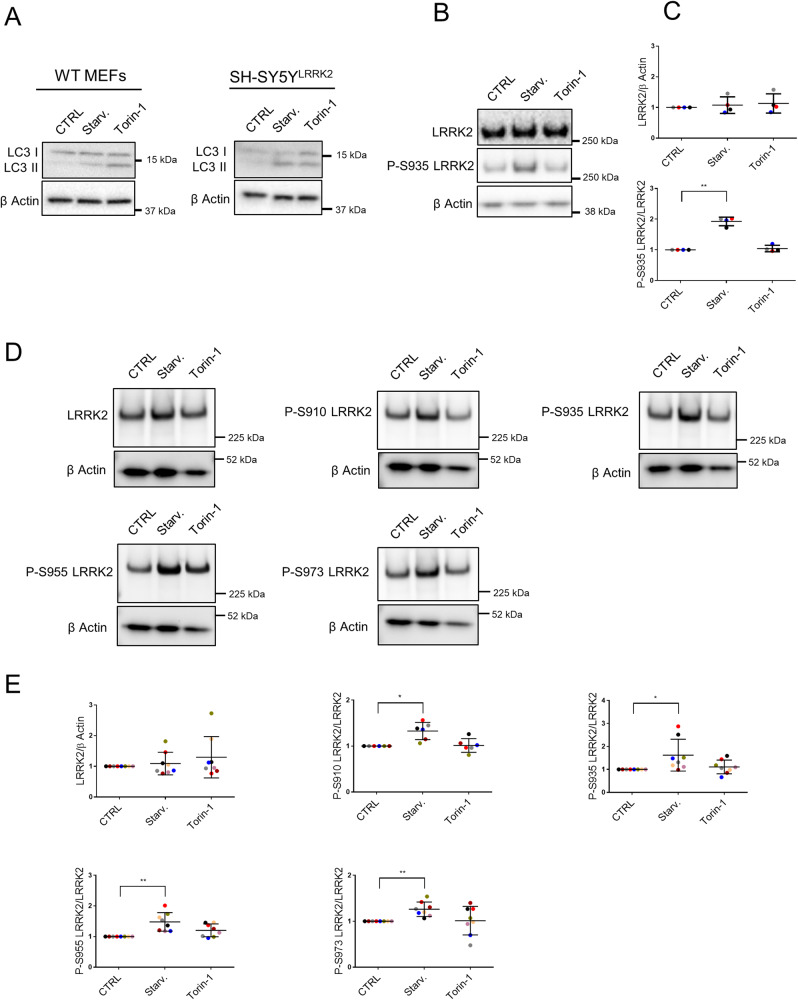
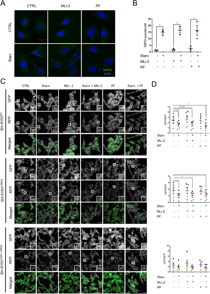
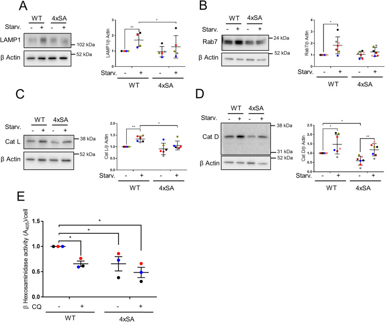
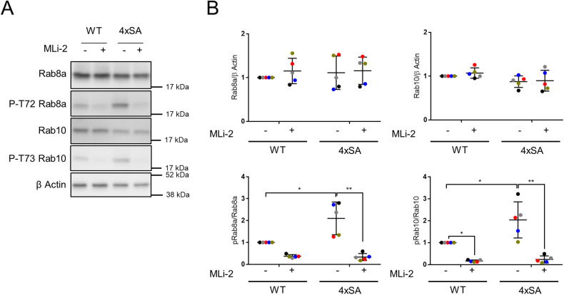
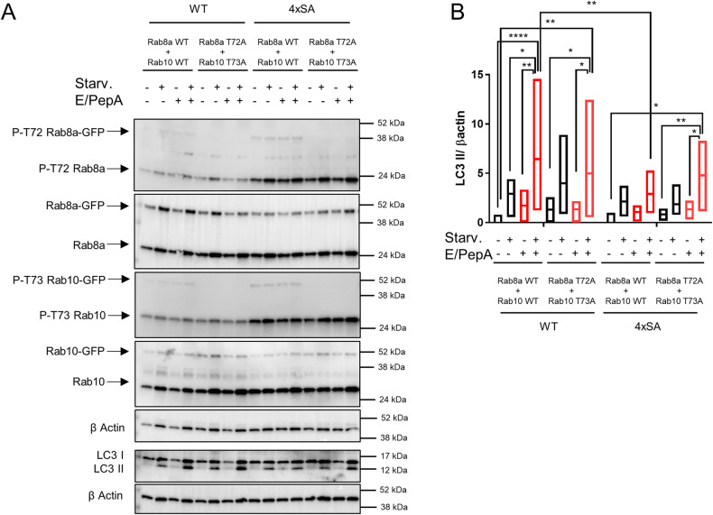
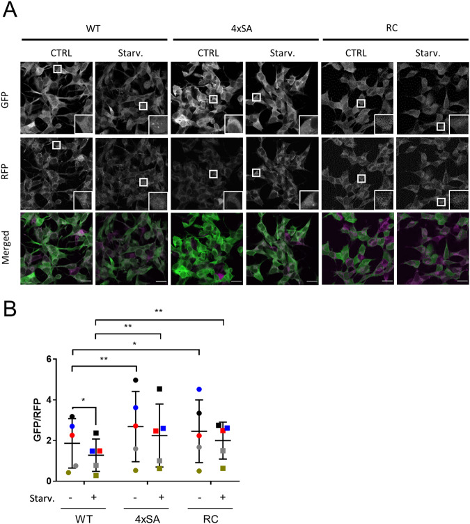
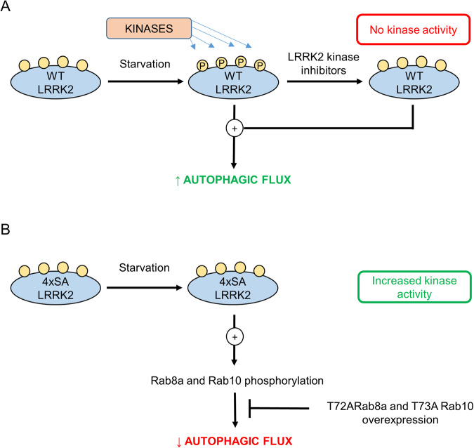
Mutations in the leucine-rich repeat kinase 2 (LRRK2) gene are the most common genetic cause of Parkinson's disease (PD), with growing importance also for Crohn's disease and cancer. LRRK2 is a large and complex protein possessing both GTPase and kinase activity. Moreover, LRRK2 activity and function can be influenced by its phosphorylation status. In this regard, many LRRK2 PD-associated mutants display decreased phosphorylation of the constitutive phosphorylation cluster S910/S935/S955/S973, but the role of these changes in phosphorylation status with respect to LRRK2 physiological functions remains unknown. Here, we propose that the S910/S935/S955/S973 phosphorylation sites act as key regulators of LRRK2-mediated autophagy under both basal and starvation conditions. We show that quadruple LRRK2 phosphomutant cells (4xSA; S910A/S935A/S955A/S973A) have impaired lysosomal functionality and fail to induce and proceed with autophagy during starvation. In contrast, treatment with the specific LRRK2 kinase inhibitors MLi-2 (100 nM) or PF-06447475 (150 nM), which also led to decreased LRRK2 phosphorylation of S910/S935/S955/S973, did not affect autophagy. In explanation, we demonstrate that the autophagy impairment due to the 4xSA LRRK2 phospho-dead mutant is driven by its enhanced LRRK2 kinase activity. We show mechanistically that this involves increased phosphorylation of LRRK2 downstream targets Rab8a and Rab10, as the autophagy impairment in 4xSA LRRK2 cells is counteracted by expression of phosphorylation-deficient mutants T72A Rab8a and T73A Rab10. Similarly, reduced autophagy and decreased LRRK2 phosphorylation at the constitutive sites were observed in cells expressing the pathological R1441C LRRK2 PD mutant, which also displays increased kinase activity. These data underscore the relation between LRRK2 phosphorylation at its constitutive sites and the importance of increased LRRK2 kinase activity in autophagy regulation and PD pathology.


Conflict of interest statement

The authors declare no competing interests.

Figures

Fig. 1
Fig. 1. LRRK2 phosphorylation increases during starvation-induced autophagy.
WT MEFs and SH-SY5YLRRK2 cells were treated for 2 h with DMSO (CTRL), starvation medium (Starv.), or Torin-1 (50 nM). Representative western blots showing A LC3 I and II levels (N = 3 independent experiments) in each cell line and B LRRK2 expression levels and phosphorylation at S935 in WT MEFs (N = 4 independent experiments). C Quantification of LRRK2 expression and S935 phosphorylation levels presented in (B). D Representative western blots showing LRRK2 expression levels and phosphorylation at S910/S935/S955/S973 in SH-SY5YLRRK2 cells (N = 6–8 independent experiments). E Quantification of LRRK2 expression and phosphorylation levels at S910/S935/S955/S973 presented in (D). β actin was used on all blots as a loading control. All graphs are presented as plots of individual data points with mean ± SD, with each experiment marked in a different color and normalized to a control condition (CTRL). Statistical analysis was performed using a Repeated Measures one-way ANOVA with Dunnett’s multiple comparisons post-test (*P < 0.05; **P < 0.001).
Fig. 2
Fig. 2. Overexpression of WT LRRK2 but not 4xSA LRRK2 phosphomutant stimulates starvation-induced autophagy.
A, B SH-SY5YEV, SH-SY5YLRRK2, and SH-SY5Y4xSA LRRK2 cells remained untreated (CTRL) or were treated for 2 h with starvation medium (Starv.). A Representative images showing WIPI2 puncta from N = 4 independent experiments with at least 1200 cells analysed per condition. Scale bar: 20 μm. B Quantification of the WIPI2 puncta per cell shown in (A). The means of all experiments ± SEM are shown with each experiment marked in a different color. For statistical analysis, a Repeated Measures two-way ANOVA with Sidak’s multiple comparisons post-test (*P < 0.05; **P < 0.01; ***P < 0.001) was used. C, D SH-SY5YEV, SH-SY5YLRRK2, and SH-SY5Y4xSA LRRK2 cells were treated for 2 h with DMSO or starvation medium (Starv.) in the absence or presence of the lysosomal protease inhibitors E64d and pepstatin A (E/PepA; 10 μM each). C Representative western blot showing LC3 I and II levels (N = 4 independent experiments). β actin was used as a loading control. D Quantification of the LC3 II levels presented in (C) shown as a plot of the individual data points with mean ± SD with each experiment marked in a different color and normalized to the empty vector CTRL (no starvation medium, no protease inhibitors). Repeated Measures two-way ANOVA with Sidak’s multiple comparisons post-test (*P < 0.05; **P < 0.01; ***P < 0.001; ****P < 0.0001) was used for statistical analysis. E, F MEFEV, MEFhLRRK2, and MEF4xSA hLRRK2 cells remained untreated (CTRL) or were treated with starvation medium (Starv.) for 1, 2, 3 and 4 h. E Representative western blot showing LC3 levels with β actin as loading control (N = 3 independent experiments). F Quantification of the LC3 II levels during the starvation time course presented in (E) shown as mean ± SD (N = 3 independent experiments) and normalized to the empty vector CTRL (no starvation medium). Repeated Measures two-way ANOVA with Sidak’s multiple comparisons post-test was used for statistical analysis: **P < 0.01 indicates differences for MEFhLRRK2 cells only; €€P < 0.01 marks the difference between MEFhLRRK2 and MEFEV cells; $$P < 0.01 and $$$P < 0.001 marks the difference between MEFhLRRK2 and MEF4xSA hLRRK2 cells.
Fig. 3
Fig. 3. LRRK2 kinase inhibition does neither impact the early stages of autophagy nor autophagic degradation.
A Representative images showing WIPI2 puncta (green) and nuclei stained with DAPI (blue) in WT MEFs treated for 2 h with DMSO (CTRL) or starvation medium (Starv.), in the absence or presence of the LRRK2 kinase inhibitors MLi-2 (100 nM) or PF-06447475 (PF, 150 nM). N = 3 independent experiments with at least 500 cells analysed per condition. Scale bar = 20 μm. B Quantification of the WIPI2 puncta per cell shown in (A). The means of each experiment ± SEM are shown; Repeated measures two-way ANOVA with Sidak’s multiple comparisons post-test (*P < 0.05) was used for statistical analysis. C, D SH-SY5YEV, SH-SY5YLRRK2, and SH-SY5Y4xSA LRRK2 cells expressing the GFP-LC3-RFP-LC3ΔG construct were treated for 4 h with DMSO (CTRL) or starvation medium (Starv.), in the absence or presence of MLi-2 (100 nM) or of PF-06447475 (PF, 150 nM). C Representative images of GFP-LC3-RFP-LC3ΔG expression are shown; scale bar = 20 μm. D Quantification of the autophagic degradation process, measured as the GFP/RFP ratio and presented as a plot of individual data points with mean ± SEM. From top to bottom, the graphs correspond to the three types of cell (SH-SY5YEV, SH-SY5YLRRK2, and SH-SY5Y4xSA LRRK2) as presented in (C); N = 5 independent experiments, each involving 6 technical replicates. Statistical analysis was performed using a Repeated Measures two-way ANOVA with Sidak’s multiple comparisons post-test (*P < 0.05).
Fig. 4
Fig. 4. Levels of lysosomal/endosomal markers and enzymes are lower in SH-SY5Y4xSA LRRK2 than in SH-SY5YLRRK2 cells.
AD SH-SY5YLRRK2 and SH-SY5Y4xSA LRRK2 cells remained untreated (CTRL) or were treated for 2 h with starvation medium (Starv.). Representative western blots showing the levels of (A) LAMP1 (N = 5 independent experiments), B Rab7 (N = 7 independent experiments), C Cathepsin L (Cat L) (N = 6 independent experiments), and D Cathepsin D (Cat D) (N = 7 independent experiments). β actin was used as a loading control. The graphs show the quantification of resp. LAMP1, Rab7, Cat L, and Cat D levels relative to the loading control, presented as a plot of individual data points with mean ± SD with each experiment marked in a different color and normalized to the control condition (WT LRRK2, no starvation); Repeated Measures two-way ANOVA with Sidak’s multiple comparisons post-test (*P < 0.05; **P < 0.01). E Graph showing the activity of β-hexosaminidase measured in SH-SY5YLRRK2 and SH-SY5Y4xSA LRRK2 cells treated for 2 h with or without chloroquine (CQ, 10 μM). The graph is presented as individual data points with mean ± SEM normalized to the control condition (WT LRRK2, no CQ); N = 3 independent experiments with 3 technical replicates per experiment. Statistical significance is shown in comparison to the control condition; Repeated Measures two-way ANOVA with Dunnett’s multiple comparisons post-test (*P < 0.05).
Fig. 5
Fig. 5. SH-SY5Y4xSA LRRK2 cells have increased LRRK2 kinase activity towards Rab8a and Rab10.
SH-SY5YLRRK2 and SH-SY5Y4xSA LRRK2 were treated for 2 h with DMSO or with the LRRK2 kinase inhibitor MLi-2 (100 nM). A Representative western blots showing the levels of total Rab8a, total Rab10, and the phosphorylation levels of Rab8a at T72 and of Rab10 at T73 (N = 5 independent experiments). β actin was used as a sample integrity control. B Quantification of the levels of total and phosphorylated Rab8a and Rab10 levels presented in (A). Graphs are presented as plots of individual data points with mean ± SD, with each experiment marked in a different color and normalized to the control condition (WT LRRK2, no MLi-2). Statistical significance is shown in comparison to the control condition (SH-SY5YLRRK2 cells in the absence of MLi-2 treatment); statistical analysis was performed using a Repeated Measures two-way ANOVA with Sidak’s multiple comparisons post-test (*P < 0.05; **P < 0.01).
Fig. 6
Fig. 6. Ectopic expression of phosphorylation-deficient T72A Rab8a and T73A Rab10 restores starvation-induced autophagic flux in SH-SY5Y4xSA LRRK2 cells.
SH-SY5YLRRK2 and SH-SY5Y4xSA LRRK2 cells were co-transfected with either pDEST53-GFP-Rab8a together with pDEST53-GFP-Rab10 or with pDEST53-GFP-Rab8aT72A together with pDEST53-GFP-Rab10T73A. Cells were treated for 2 h with DMSO or starvation medium (Starv.) in the absence or presence of lysosomal proteases inhibitors E64d and pepstatin A (E/PepA; 10 μM each). A Representative western blots show the levels of total Rab8a, total Rab10, and the levels of P-T72 Rab8a and of P-73 Rab10 as well as of LC3 I and LC3 II (N = 3–5 independent experiments). β actin was used as a sample integrity control. B Quantification of the LC3 II levels presented in (A) normalized to the empty vector CTRL (no starvation medium, no protease inhibitors). Floating bars show the minimum and maximum values for each condition, with the line indicating the mean (N = 5 independent experiments). Samples treated with E64d and pepstatin A are shown in red. Repeated Measures two-way ANOVA with Sidak’s multiple comparisons post-test (*P < 0.05; **P < 0.01; ****P < 0.0001) was used for statistical analysis.
Fig. 7
Fig. 7. SH-SY5Y4xSA LRRK2 and SH-SY5YR1441C LRRK2 cells demonstrate a similarly impaired basal and starvation-induced autophagic flux.
SH-SY5YLRRK2, SH-SY5Y4xSA LRRK2, and SH-SY5YR1441C LRRK2 cells expressing the GFP-LC3-RFP-LC3ΔG construct were analysed for autophagic degradation by measuring the GFP/RFP ratio. The cells were treated with DMSO (CTRL) or with starvation medium (Starv.) for 2 h. A Representative image showing GFP-LC3-RFP-LC3ΔG expression; scale bar = 20 μm. B Quantification of autophagic degradation measured as GFP/RFP ratio is presented as a plot of individual data points with mean ± SEM; N = 5 independent experiments each involving three to six technical replicates. Statistical analysis was performed using a Repeated Measures two-way ANOVA with Sidak’s multiple comparisons post-test (*P < 0.05; **P < 0.01). Please note that the data shown in (B) form part of the data set also presented in Fig. 3D.
Fig. 8
Fig. 8. Model of autophagy regulation by LRRK2 phosphorylation at S910/S935/S955/S973 during starvation.
A During starvation, WT LRRK2 is extensively phosphorylated at S910/S935/S955/S973, stimulating the autophagic process. Treatment with the LRRK2 kinase inhibitors MLi-2 or PF-06447475 inhibits the LRRK2 kinase activity and leads to S910/S935/S955/S973 dephosphorylation. B Substitution of the serines with alanines (S910A/S935A/S955A/S973A; 4xSA LRRK2) prohibits their phosphorylation, also under starvation conditions. However, in contrast to what happens during treatment with LRRK2 inhibitors, 4xSA LRRK2 is characterized by increased kinase activity. Due to the resulting increased downstream phosphorylation of Rab8a and Rab10, autophagic flux is impaired. The latter can be rescued by overexpression of phospho-dead mutants T72A Rab8a and T73A Rab10. Color code: green, stimulation; red, inhibition.

References

    1. Lewis PA. The function of ROCO proteins in health and disease. Biol Cell. 2009;101:183–91. doi: 10.1042/BC20080053. - DOI - PubMed
    1. Hui KY, Fernandez-Hernandez H, Hu J, Schaffner A, Pankratz N, Hsu NY, et al. Functional variants in the LRRK2 gene confer shared effects on risk for Crohn’s disease and Parkinson’s disease. Sci Transl Med. 2018;10:eaai7795. doi: 10.1126/scitranslmed.aai7795. - DOI - PMC - PubMed
    1. Saunders-Pullman R, Barrett MJ, Stanley KM, San Luciano M, Shanker V, Severt L, et al. LRRK2 G2019S mutations are associated with an increased cancer risk in Parkinson disease. Mov Disord. 2010;25:2536–41. doi: 10.1002/mds.23314. - DOI - PMC - PubMed
    1. Paisán-Ruiz C, Jain S, Evans EW, Gilks WP, Simon J, van der Brug M, et al. Cloning of the gene containing mutations that cause PARK8-linked Parkinson’s disease. Neuron. 2004;44:595–600. doi: 10.1016/j.neuron.2004.10.023. - DOI - PubMed
    1. Zimprich A, Biskup S, Leitner P, Lichtner P, Farrer M, Lincoln S, et al. Mutations in LRRK2 cause autosomal-dominant parkinsonism with pleomorphic pathology. Neuron. 2004;44:601–7. doi: 10.1016/j.neuron.2004.11.005. - DOI - PubMed

Publication types

MeSH terms

Substances

LinkOut - more resources