Multicenter Study
. 2023 Nov;43(11):1392-1397.
doi: 10.1038/s41372-023-01723-3. Epub 2023 Jul 15.

Neonatal brain MRI and short-term outcomes after acute provoked seizures

Affiliations
Multicenter Study

Neonatal brain MRI and short-term outcomes after acute provoked seizures

Yi Li et al. J Perinatol. 2023 Nov.

Abstract

Objective: We investigated how diagnosis and injury location on neonatal brain MRI following onset of acute provoked seizures was associated with short term outcome.

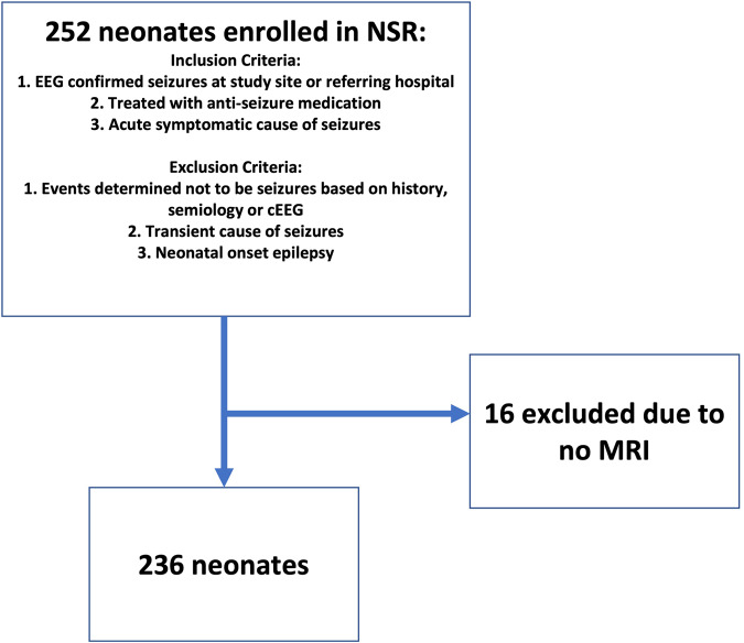
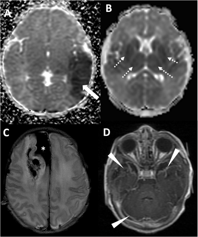
Study design: A multicenter cohort of neonates with acute provoked seizures enrolled in the Neonatal Seizure Registry. MRIs were centrally evaluated by a neuroradiologist for location of injury and radiologic diagnosis. Clinical outcomes were determined by chart review. Multivariate logistic regression was used to examine the association between MRI findings and outcomes.

Results: Among 236 newborns with MRI at median age 4 days (IQR 3-8), 91% had abnormal MRI. Radiologic diagnoses of intracranial hemorrhage (OR 3.2 [1.6-6.5], p < 0.001) and hypoxic-ischemic encephalopathy (OR 2.7 [1.4-5.4], p < 0.003) were associated with high seizure burden. Radiologic signs of intracranial infection were associated with abnormal neurologic examination at discharge (OR 3.9 [1.3-11.6], p < 0.01).

Conclusion: Findings on initial MRI can help with expectant counseling on short-term outcomes following acute provoked neonatal seizures.


Conflict of interest statement

RS is the only author with relevant disclosures, including: Associate Editor for Neurology; consultant for the Epilepsy Study Consortium; and receives royalties from UpToDate for authorship of topics related to neonatal seizures. The other authors have no relevant disclosures or conflicts of interest.

Figures

Fig. 1
Fig. 1. Inclusion and exclusion criteria for the study.
After exclusion of 16 neonates with no MRI, the final cohort consisted of 236 neonates.
Fig. 2
Fig. 2. Examples of injury based on location.
A Axial T2 weighted imaging demonstrating cortical injury in the setting of arterial ischemic stroke, with missing cortical ribbon (white arrow) in area of edematous, injured cortex. B Axial ADC map demonstrating reduced diffusion in the deep gray nuclei signifying acute injury (arrowheads).
Fig. 3
Fig. 3. Radiologic diagnoses in the cohort of patients with acute symptomatic seizures.
A Axial ADC map demonstrating arterial ischemic stroke in the left middle cerebral artery territory (white arrow). B Axial ADC map demonstrating reduced diffusion in the deep gray nuclei and subcortical white matter, compatible with hypoxic ischemic injury. C Axial T2 weighted imaging demonstrating large subpial hemorrhage in the medial right frontal lobe (asterisk) with associated hemorrhagic injury of the right frontal lobe. D Axial T1-weighted post-contrast imaging demonstrating leptomeningeal enhancement (arrowheads) in the setting of neonatal meningitis/intracranial infection.

References

    1. Glass HC, Grinspan ZM, Shellhaas RA. Outcomes after acute symptomatic seizures in neonates. Semin Fetal Neonatal Med. 2018;23:218–22. doi: 10.1016/j.siny.2018.02.001. - DOI - PubMed
    1. Glass HC, Pham TN, Danielsen B, Towner D, Glidden D, Wu YW. Antenatal and intrapartum risk factors for seizures in term newborns: a population-based study, California 1998-2002. J Pediatr. 2009;154:24–8. doi: 10.1016/j.jpeds.2008.07.008. - DOI - PMC - PubMed
    1. Glass HC, Shellhaas RA, Wusthoff CJ, Chang T, Abend NS, Chu CJ, et al. Contemporary profile of seizures in neonates: a prospective cohort study. J Pediatr. 2016;174:98–103.e1. doi: 10.1016/j.jpeds.2016.03.035. - DOI - PMC - PubMed
    1. Glass HC, Hong KJ, Rogers EE, Jeremy RJ, Bonifacio SL, Sullivan JE, et al. Risk factors for epilepsy in children with neonatal encephalopathy. Pediatr Res. 2011;70:535–40. doi: 10.1203/PDR.0b013e31822f24c7. - DOI - PMC - PubMed
    1. McDonough TL, Paolicchi JM, Heier LA, Das N, Engel M, Perlman JM, et al. Prediction of future epilepsy in neonates with hypoxic-ischemic encephalopathy who received selective head cooling. J Child Neurol. 2017;32:630–7. doi: 10.1177/0883073817698628. - DOI - PubMed

Publication types