. 2023 Jul 25;42(7):112777.
doi: 10.1016/j.celrep.2023.112777. Epub 2023 Jul 16.

VEGFR3 is required for button junction formation in lymphatic vessels

Affiliations

VEGFR3 is required for button junction formation in lymphatic vessels

Melanie Jannaway et al. Cell Rep. .

Abstract

Lymphatic capillaries develop discontinuous cell-cell junctions that permit the absorption of large macromolecules, chylomicrons, and fluid from the interstitium. While excessive vascular endothelial growth factor 2 (VEGFR2) signaling can remodel and seal these junctions, whether and how VEGFR3 can alter lymphatic junctions remains incompletely understood. Here, we use lymphatic-specific Flt4 knockout mice to investigate VEGFR3 signaling in lymphatic junctions. We show that loss of Flt4 prevents specialized button junction formation in multiple tissues and impairs interstitial absorption. Knockdown of FLT4 in human lymphatic endothelial cells results in impaired NOTCH1 expression and activation, and overexpression of the NOTCH1 intracellular domain in Flt4 knockout vessels rescues the formation of button junctions and absorption of interstitial molecules. Together, our data reveal a requirement for VEGFR3 and NOTCH1 signaling in the development of button junctions during postnatal development and may hold clinical relevance to lymphatic diseases with impaired VEGFR3 signaling.

Keywords: CP: Developmental biology; FLT4; Milroy; NOTCH; VEGFR3; button; junctions; lymphatic; lymphedema; zipper.


Conflict of interest statement

Declaration of interests The authors declare no competing interest.

Figures
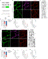
Figure 1.
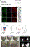
Figure 1.. Lymphatic-specific deletion of Flt4 prevents the appearance of buttons in the diaphragm and ear skin
(A) Tamoxifen injection procedure for deletion of Flt4 to assess button formation. (B) P21 diaphragms immunostained for GFP (green), VEGFR3 (magenta), and VE-cadherin (red). (C) Higher magnification images of lymphatic vessel junction morphology. (D) Quantification of button, intermediate, and zipper junctions in Flt4fl/fl (N = 7 mice; n = 11 fields of view [FOVs]) and Flt4iLECKO (N = 6 mice; n = 12 FOVs) mice. (E–G) A breakdown of the graph in (D) for the indicated junction types. (H) P21 ear skin was stained for GFP (green), VEGFR3 (magenta), and VE-cadherin (red). (I) Higher magnification of VE-cadherin at intercellular junctions. (J) Junction morphology quantification in Flt4fl/fl (N = 5 mice; n = 11 FOVs) and Flt4iLECKO (N = 6; n = 12 FOVs) mice. (K–M) Individual graphs for each of the three junction types presented in (J). Two-way ANOVA with Sidak’s test. ****p < 0.0001 for buttons; †††p < 0.001 for intermediate; ‡‡‡‡p < 0.0001 for zippers; ns, non-significant for intermediate. All data are presented as mean ± SD. Scale bar in (B) and (H) represents 50 μm and in (C) and (I) represents 10 μm. FOVs, fields of view.
Figure 2.
Figure 2.. VEGFR3 is not required for button maintenance
(A) Tamoxifen injection procedure to delete Flt4 after buttons have formed to investigate button maintenance. (B) P35 diaphragms were immunostained for GFP (green), VEGFR3 (magenta), and VE-cadherin (red). (C) Higher magnification of junction phenotype in lymphatic capillaries. (D) Quantification of junction phenotype in Flt4fl/fl (N = 5 mice; n = 11 FOVs) and Flt4iLECKO (N = 5 mice; n = 8 FOVs) mice. (E–G) Individual graphs of each junction type presented in (D). Two-way ANOVA with Sidak’s test. ns, non-significant. All data are presented as mean ± SD. Scale bar in (B) represents 50 μm, and in (C), it represents 10 μm. FOVs, fields of view.
Figure 3.
Figure 3.. FLT4 knockdown inhibits Notch signaling and expression
(A) qRT-PCR analysis for members of the Notch signaling pathway and other putative signaling pathways associated with VEGFR3 in cultured human dermal lymphatic endothelial cells. (B) Western blot for VEGFR3, pVEGFR2, VEGFR2, cleaved NOTCH1, cleaved NOTCH4, VE-cadherin, and DLL4 to assess activation of these signaling pathways. (C) Quantification of the western blot data in (B). **p < 0.01; ***p < 0.001; ****p < 0.0001; ns, non-significant. Student’s unpaired t test. All data are presented as mean ± SEM. All experiments were performed n = 3–4 times.
Figure 4.
Figure 4.. Overexpression of the NOTCH1 intracellular domain (NICD1) rescues button formation and lymphatic function in the absence of VEGFR3
(A) Tamoxifen procedure for deletion of Flt4 and induction of constitutive NICD1 expression. (B) Lymphatic vessels in P21 diaphragms were immunostained for GFP (green), VEGFR3 (magenta), and VE-cadherin (red). (C) Higher magnification images of VE-cadherin immunostaining at lymphatic endothelial intercellular junctions. (D) Quantification of the three different junction types in Flt4fl/fl;R26NICD1 (N = 5 mice; n = 15 FOVs), Flt4iLECKO (N = 4 mice; n = 11 FOVs), and Flt4iLECKO;R26NICD1 (N = 4 mice; n = 11 FOVs) mice. (E–G) Individual graphs of button, intermediate and zipper junctions presented in (D). (H) Evans blue dye was injected into the ears of Flt4fl/fl;R26NICD1, Flt4iLECKO, and Flt4iLECKO;R26NICD1 mice at P21, and dye uptake into the lymphatic vasculature was imaged. (I) BSA conjugated to Alexa 790 was injected into P24 mouse ears and imaged. Two-way ANOVA with Sidak’s post hoc test. ****p < 0.0001 buttons; ‡‡‡‡p < 0.0001 zippers; ns, non-significant. All data are presented as mean ± SD. Scale bar in (B) represents 50 μm, in (C) represents 10 μm, in (H) represents 500 μm, and in (I) represents 1 mm. FOVs, fields of view.

References

    1. Brouillard P, Witte MH, Erickson RP, Damstra RJ, Becker C, Quéré I, and Vikkula M (2021). Primary lymphoedema. Nat. Rev. Dis. Primers 7, 77. 10.1038/s41572-021-00309-7. - DOI - PubMed
    1. Iyer D, Jannaway M, Yang Y, and P Scallan J (2020). Lymphatic valves and lymph flow in cancer-related lymphedema. Cancers 12, 2297. 10.3390/cancers12082297. - DOI - PMC - PubMed
    1. Baluk P, Fuxe J, Hashizume H, Romano T, Lashnits E, Butz S, Vestweber D, Corada M, Molendini C, Dejana E, and McDonald DM (2007). Functionally specialized junctions between endothelial cells of lymphatic vessels. J. Exp. Med 204, 2349–2362. 10.1084/jem.20062596. - DOI - PMC - PubMed
    1. Yao LC, Baluk P, Srinivasan RS, Oliver G, and McDonald DM (2012). Plasticity of button-like junctions in the endothelium of airway lymphatics in development and inflammation. Am. J. Pathol 180, 2561–2575. 10.1016/j.ajpath.2012.02.019. - DOI - PMC - PubMed
    1. Zheng W, Nurmi H, Appak S, Sabine A, Bovay E, Korhonen EA, Orsenigo F, Lohela M, D’Amico G, Holopainen T, et al. (2014). Angiopoietin 2 regulates the transformation and integrity of lymphatic endothelial cell junctions. Genes Dev 28, 1592–1603. 10.1101/gad.237677.114. - DOI - PMC - PubMed

Publication types

MeSH terms

Substances

LinkOut - more resources