. 2024 Jun;309(6):2505-2513.
doi: 10.1007/s00404-023-07133-2. Epub 2023 Jul 16.

Controlled prospective study on ultrasound simulation training in fetal echocardiography: FESIM II

Affiliations

Controlled prospective study on ultrasound simulation training in fetal echocardiography: FESIM II

Paul Janzing et al. Arch Gynecol Obstet. 2024 Jun.

Abstract

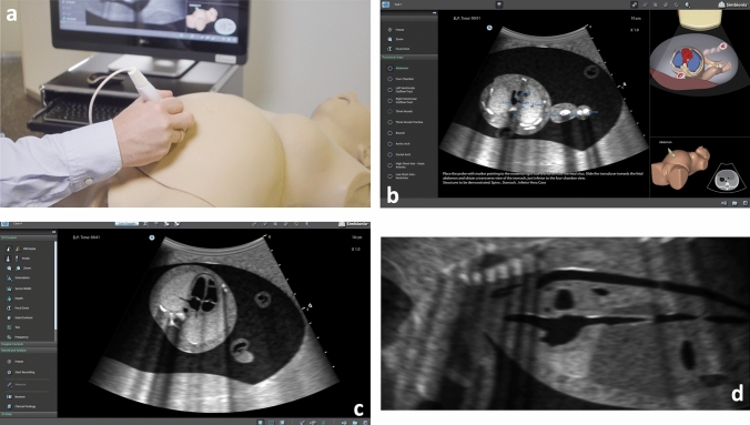
Purpose: To analyze the learning curves of ultrasound novices in fetal echocardiography during structured simulation-based ultrasound training (SIM-UT) including a virtual, randomly moving fetus.

Methods: 11 medical students with minimal (< 10 h) prior obstetric ultrasound experience underwent 12 h of structured fetal echocardiography SIM-UT in individual hands-on sessions during a 6-week training program. Their learning progress was assessed with standardized tests after 2, 4, and 6 weeks of SIM-UT. Participants were asked to obtain 11 fetal echocardiography standard planes (in accordance with ISUOG and AHA guidelines) as quickly as possible. All tests were carried out under real life, examination-like conditions on a healthy, randomly moving fetus. Subsequently, we analyzed the rate of correctly obtained images and the total time to completion (TTC). As reference groups, 10 Ob/Gyn physicians (median of 750 previously performed Ob/Gyn scans) and 10 fetal echocardiography experts (median of 15,000 previously performed Ob/Gyn scans) were examined with the same standardized tests.

Results: The students showed a consistent and steady improvement of their ultrasound performance during the training program. After 2 weeks, they were able to obtain > 95% of the standard planes correctly. After 6 weeks, they were significantly faster than the physician group (p < 0.001) and no longer significantly slower than the expert group (p = 0.944).

Conclusion: SIM-UT is highly effective to learn fetal echocardiography. Regarding the acquisition of the AHA/ISUOG fetal echocardiography standard planes, the students were able to reach the same skill level as the expert group within 6 weeks.

Keywords: Congenital heart disease; Fetal echocardiography; Prenatal detection rate; Simulation-based ultrasound training; Ultrasound didactics; Ultrasound simulation training.


Conflict of interest statement

The authors declare that no funds, grants, or other support were received during the preparation of this manuscript. The authors have no financial or non-financial interests to disclose.

Figures

Fig. 1
Fig. 1
The US Mentor ultrasound simulator: a Setup of the simulator. b Screen in “Task mode” (with aids), three vessel view. On the left side: the desired standard plane can be selected. Center: B-mode image including anatomic labels. Below the B-mode image: written instructions on how to obtain the correct standard plane. Right top corner: 3D model of the fetus demonstrating the current plane. Right bottom corner: Position of the probe on the mannequin. c Screen during a standardized examination, four chamber view. No aids are given by the simulator. Left side: “Knobology” of the ultrasound machine, e.g., adjustment of the depth or focus zone. d A bicaval plane assessed as incorrect by the simulator that was later rated as correct (Grade 1−) by the expert panel
Fig. 2
Fig. 2
Boxplot showing the distribution of total time to completion (TTC) in seconds of all standard planes by students after 2, 4, and 6 weeks training time compared to physician and expert groups. #Significant difference within groups using Wilcoxon test (p < 0.05), *significant difference between groups using Mann–Whitney U test (p < 0.05)
Fig. 3
Fig. 3
Boxplot showing the distribution of time to completion [s] of single planes between students (after 6 weeks training), physicians and experts. Groups were compared by Mann–Whitney U-test. A p < 0.05 was considered as significant. Standard views: Abd (transverse abdominal), 4CV (four chamber), LVOT (left ventricular outflow tract), RVOT (right ventricular outflow tract), 3V (three vessels), 3VT (three vessels trachea), Bic (bicaval), Ao (aortic arch), Duc (ductal arch), HSX (high short axis), LSX (low short axis). #Significant difference between students and physicians, *significant difference between students and experts, §significant difference between physicians and experts

References

    1. Lindinger A, Schwedler G, Hense HW. Prevalence of congenital heart defects in newborns in Germany: results of the first registration year of the PAN study (July 2006 to June 2007) Klin Padiatr. 2010;222:321–326. doi: 10.1055/s-0030-1254155. - DOI - PubMed
    1. van der Linde D, Konings EEM, Slager MA, et al. Birth prevalence of congenital heart disease worldwide: a systematic review and meta-analysis. J Am Coll Cardiol. 2011;58:2241–2247. doi: 10.1016/J.JACC.2011.08.025. - DOI - PubMed
    1. Morris SA, Ethen MK, Penny DJ, et al. Prenatal diagnosis, birth location, surgical center, and neonatal mortality in infants with hypoplastic left heart syndrome. Circulation. 2014;129:285–292. doi: 10.1161/CIRCULATIONAHA.113.003711. - DOI - PMC - PubMed
    1. Tworetzky W, McElhinney DB, Reddy VM, et al. Improved surgical outcome after fetal diagnosis of hypoplastic left heart syndrome. Circulation. 2001;103:1269–1273. doi: 10.1161/01.CIR.103.9.1269. - DOI - PubMed
    1. Mahle WT, Clancy RR, McGaurn SP, et al. Impact of prenatal diagnosis on survival and early neurologic morbidity in neonates with the hypoplastic left heart syndrome. Pediatrics. 2001;107:1277–1282. doi: 10.1542/peds.107.6.1277. - DOI - PubMed