Review
. 2023 Sep;45(9):e2300068.
doi: 10.1002/bies.202300068. Epub 2023 Jul 16.

A short story on how chromophore is hydrolyzed from rhodopsin for recycling

Affiliations
Review

A short story on how chromophore is hydrolyzed from rhodopsin for recycling

John D Hong et al. Bioessays. 2023 Sep.

Abstract

The photocycle of visual opsins is essential to maintain the light sensitivity of the retina. The early physical observations of the rhodopsin photocycle by Böll and Kühne in the 1870s inspired over a century's worth of investigations on rhodopsin biochemistry. A single photon isomerizes the Schiff-base linked 11-cis-retinylidene chromophore of rhodopsin, converting it to the all-trans agonist to elicit phototransduction through photoactivated rhodopsin (Rho*). Schiff base hydrolysis of the agonist is a key step in the photocycle, not only diminishing ongoing phototransduction but also allowing for entry and binding of fresh 11-cis chromophore to regenerate the rhodopsin pigment and maintain light sensitivity. Many challenges have been encountered in measuring the rate of this hydrolysis, but recent advancements have facilitated studies of the hydrolysis within the native membrane environment of rhodopsin. These techniques can now be applied to study hydrolysis of agonist in other opsin proteins that mediate phototransduction or chromophore turnover. In this review, we discuss the progress that has been made in characterizing the rhodopsin photocycle and the journey to characterize the hydrolysis of its all-trans-retinylidene agonist.

Keywords: 11-cis-retinal; all-trans-retinal; chromophore; dark adaptation; retinal hydrolysis; retinylidene phospholipids; rhodopsin.


Conflict of interest statement

Conflict of interest: K.P. is a consultant for Polgenix Inc. J.D.H. has declared that no conflict of interest exists.

Figures

Figure 1.
Figure 1.. Three dimensional models of rhodopsin and cone opsins.
Residues surrounding the chromophore are depicted for rhodopsin and three human cone opsin pigments. The rhodopsin model is based on PDB 1U19, and the homology models of the cone opsin pigments are deposited in the PDB (identifiers 1KPN, 1KPW, and 1KPX for the blue, green, and red cone pigments, respectively) [110]. Each pigment has a characteristic absorbance maximum (λmax): rhodopsin (500 nm), red (560 nm), green (530 nm), blue (420 nm). The absorbance maximum of the protonated retinylidene Schiff base (λmax = 440 nm) undergoes a bathochromic shift by interaction of the polyene chain with key residues in the chromophore binding pockets of rhodopsin, red opsin, and green opsin; whereas for blue opsin, a hypsochromic shift occurs. Abbreviations: Ret, retinylidene; PLM, palmitoyl; NAG, N-acetylglucosamine; HTG, Heptyl 1-thio-β-D-glucopyranoside
Figure 2.
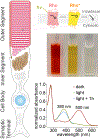
Figure 2.. Rod photoreceptor with rhodopsin photochemistry.
Rhodopsin is abundant and tightly packed in rod outer segment discs for high sensitivity to light, needed for optimal capture of photons under dim light conditions. Light induces the characteristic color changes that were observed originally by Böll and Kühne, with gradual bleaching of the red pigment of rhodopsin to the visual yellow form of Rho*, and eventually to the visual white (or colorless) form of apo-opsin with free retinal [16,58]. The broad absorbance spectrum with a λmax of 500 nm results in the apparent red color of rhodopsin. The change in absorption maximum to 380 nm from light exposure results in the apparent yellow color of Rho*. The fading of the yellow pigment is due to the hydrolysis of Rho*, releasing all-trans-retinal with a lower extinction coefficient than the deprotonated retinylidene Schiff base adducted to Rho*.
Figure 3.
Figure 3.. Rhodopsin photocycle.
The photocycle is initiated upon photoisomerization of the 11-cis-retinylidene chromophore of ground-state rhodopsin to the all-trans configuration. The substantial change in retinylidene ligand structure from the cis-to-trans configuration induces a series of conformation changes in rhodopsin. Exceedingly transient key photointermediates are formed in the process of relaxation of the protein structure to accommodate the all-trans agonist in route to the conformation of the MII active signaling state. The subsequent step of hydrolysis is essential not only to the rhodopsin photocycle but also to the visual cycle. Hydrolysis of the retinylidene Schiff base in MII releases all-trans-retinal, resulting in apo-opsin that can be regenerated to ground-state rhodopsin by binding fresh 11-cis-retinal from the visual cycle. MII can alternatively convert to MIII which possesses the syn-configuration of the retinylidene Schiff base, which hydrolyzes significantly slower. PDB IDs used were: bathorhodopsin, 2G87 [73]; lumirhodopsin, 2HPY [74]; MII, 3PXO [22], 3PQR [22], 2I37 [100]; opsin, 3CAP [88]; rhodopsin 1F88 [86]; 1L9H [78].
Figure 4.
Figure 4.. Mechanism of the hydrolysis of the all-trans-retinylidene agonist of Rho*.
The following diagram was adapted from Palczewski 2006 [84] and updated with mechanistic descriptions from Hong, et al. 2022 [45]. Light exposure of rhodopsin momentarily forms MI, which quickly deprotonates to form MII, the active signaling state for phototransduction. The extensive water network within rhodopsin provides an aqueous environment [116] which can mediate the hydrolysis process through MII [50,116]. The protic environment provided by water allows for quick transfer of protons as well as H-bonding interactions to help stabilize each intermediate. Nucleophilic attack by water on C15 of the retinylidene Schiff base forms an intermediate stabilized by H-bonding interactions with water [50]. A series of proton transfers mediated by the protic environment of water produces the carbinol-ammonium intermediate, which is primed for the completion of hydrolysis, with the protonated Lys residue serving as a favorable leaving group. The elimination of the protonated Lys residue leads to formation of the all-trans-retinal hydrolysis product. The mechanistic route as well as the protonation states of E113 and E181 shown for MII were based on quantum chemical calculations [45]. The actual protonation state of E181 is unclear, with different conclusions having been reported in previous studies; however, more recent studies have corroborated a deprotonated E181 [33,62,89,101,125]. If and how E181 protonation status affects the hydrolysis mechanism requires further experimental investigation to potentially enrich the description above; nevertheless, the presence of E181 in extracellular loop 2 serves to stabilize the Schiff-base-bound chromophore against hydrolysis [124].
Figure 5.
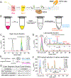
Figure 5.. Characterizing visual cycle kinetics in the rod outer segment
(A) Schematic diagram depicting the visual cycle processes of the ROS, initiated upon light exposure. In black: retinal, in blue: retinol. (B) Experimental diagram depicting iPrOH/NaBH4 treatment of ROS membranes for separation of the protein-precipitate pellet, which is proteolyzed and analyzed by LC-MS/MS for measurement of opsin-bound retinal; in turn, the lipid soluble supernatant is analyzed by LC-MS/MS to measure retinal produced by Schiff base hydrolysis in Rho* hydrolysis, and subsequent N-retinylidene-PE adducts. (C) Progressive chromatographic traces reflecting the status of the retinylidene Schiff base bound to rhodopsin, as a function of time after light exposure. The 11-cis to all-trans photoisomerization is captured by the pronase digestion of iPrOH/NaBH4-treated ROS membranes. The Nε-all-trans-retinyl-Lys signal decreases over time, corresponding to the hydrolysis of the all-trans-retinylidene Schiff base of Rho*. (D) A corresponding increase in free all-trans-retinal product from Rho* hydrolysis is observed over time, as well as production of all-trans-retinylidene Schiff base adducts with phosphatidylethanolamines from the outer segment membrane. (E) Scheme depicting the tracking of NADPH-dependent reduction of retinal by outer segment RDHs as follows: deuterium-isotope labeling of retinal by NaBD4 (used in place of NaBH4) distinguishes the retinal that is yet to be reduced by RDHs. (F) Addition of NADPH to the outer segment membranes leads to reduction of retinal to retinol by native RDHs, with a corresponding gradual decline of phosphatidylethanolamine adducts. (G) Kinetics of each major process occurring after the exposure of the rod outer segment membranes to light at 20°C and at physiologic pH 7.4, showing Rho* hydrolysis as the rate limiting step of the visual cycle within the rod outer segment. Panels B-E were adapted from: Hong, J. D., Salom, D., Kochman, M. A., Kubas, A., Kiser, P. D., and Palczewski, K. (2022) Chromophore hydrolysis and release from photoactivated rhodopsin in native membranes, Proc Natl Acad Sci U S A 119, e2213911119 [45].

References

    1. Airan RD, Thompson KR, Fenno LE, Bernstein H, & Deisseroth K (2009). Temporally precise in vivo control of intracellular signalling. Nature, 458(7241), 1025–1029. doi: 10.1038/nature07926 - DOI - PubMed
    1. Anderson RE, & Maude MB (1970). Phospholipids of bovine outer segments. Biochemistry, 9(18), 3624–3628. doi: 10.1021/bi00820a019 - DOI - PubMed
    1. Andruniow T, & Olivucci M (2009). How does the relocation of internal water affect resonance Raman spectra of rhodopsin? An insight from CASSCF/Amber calculations. J Chem Theory Comput, 5(11), 3096–3104. doi: 10.1021/ct900071c - DOI - PubMed
    1. Angel TE, Chance MR, & Palczewski K (2009). Conserved waters mediate structural and functional activation of family A (rhodopsin-like) G protein-coupled receptors. Proc Natl Acad Sci U S A, 106(21), 8555–8560. doi: 10.1073/pnas.0903545106 - DOI - PMC - PubMed
    1. Angel TE, Gupta S, Jastrzebska B, Palczewski K, & Chance MR (2009). Structural waters define a functional channel mediating activation of the GPCR, rhodopsin. Proc Natl Acad Sci U S A, 106(34), 14367–14372. doi: 10.1073/pnas.0901074106 - DOI - PMC - PubMed

Publication types