. 2023 Sep:65:102816.
doi: 10.1016/j.redox.2023.102816. Epub 2023 Jul 11.

Antioxidant mitoquinone suppresses benign prostatic hyperplasia by regulating the AR-NLRP3 pathway

Affiliations

Antioxidant mitoquinone suppresses benign prostatic hyperplasia by regulating the AR-NLRP3 pathway

Bo-Ram Jin et al. Redox Biol. 2023 Sep.

Abstract

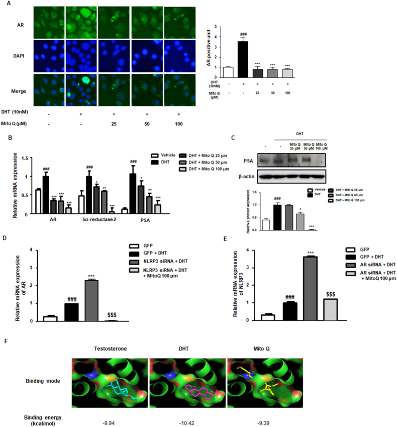
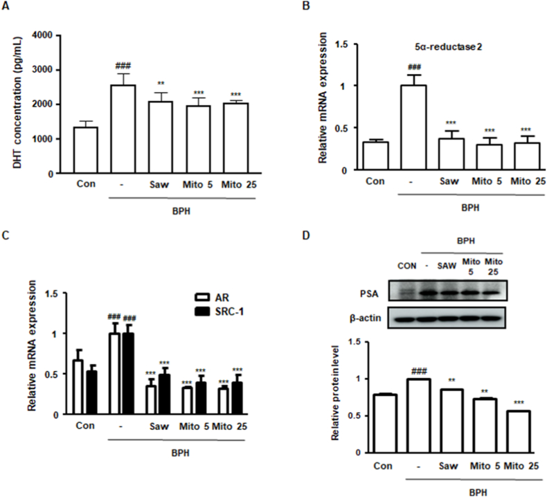
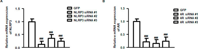
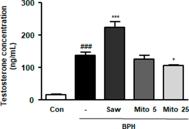
Mitoquinone (MitoQ), a mitochondria-targeted antioxidant, has been used to treat several diseases. The present study aimed to investigate the therapeutic effects of MitoQ in benign prostatic hyperplasia (BPH) models and their underlying molecular mechanisms. In this study, we determined that MitoQ inhibited dihydrotestosterone (DHT)-induced cell proliferation and mitochondrial ROS by inhibiting androgen receptor (AR) and NOD-like receptor family pyrin domain-containing 3 (NLRP3) signaling in prostate epithelial cells. Molecular modeling revealed that DHT may combine with AR and NLRP3, and that MitoQ inhibits both AR and NLRP3. AR and NLRP3 downregulation using siRNA showed the linkage among AR, NLRP3, and MitoQ. MitoQ administration alleviated pathological prostate enlargement and exerted anti-proliferative and antioxidant effects by suppressing the AR and NLRP3 signaling pathways in rats with BPH. Hence, our findings demonstrated that MitoQ is an inhibitor of NLPR3 and AR and a therapeutic agent for BPH treatment.

Keywords: Androgen receptor; Benign prostatic hyperplasia; Dihydrotestosterone; Mitoquinone; NLRP3.


Conflict of interest statement

Declaration of competing interest The authors declare that they have no known competing financial interests or personal relationships that could have appeared to influence the work reported in this paper.

Figures

Image 1
Graphical abstract
Fig. 1
Fig. 1
Antioxidant effect of MitoQ in DHT-stimulated RWPE-1 cells. (A) Molecular structure of MitoQ. (B) Viability of MitoQ-treated RWPE-1 cells at the indicated concentration for 24 h measured via the CCK-8 assay. (C) RWPE-1 cell proliferation under DHT (10 nM) stimulation and MitoQ at the indicated concentration for 24 h measured via the CCK-8 assay. (D) The expression of 8OHDG in DHT-stimulated RWPE-1 cells treated with MitoQ (25, 50, and 100 μM). (E) The mRNA expression of HO-1 and GPX-1 in DHT-stimulated RWPE-1 cells treated with MitoQ (25, 50, and 100 μM). The results are expressed as means ± SD (n = 3). ###p < 0.001 vs. vehicle group; **p < 0.01, ***p < 0.001 vs. DHT-stimulated group. (F) The manifestation of MitoTracker™ and MitoSOX™ in DHT-stimulated MitoQ-treated RWPE-1 cells (100 μM); 0.6% H2O2 was used as positive control. (G) Catalase and SOD2 protein expression in DHT-stimulated RWPE-1 cells treated with MitoQ (25, 50, and 100 μM). The results are expressed as means ± SD (n = 3). ###p < 0.001 vs. vehicle group; **p < 0.01, ***p < 0.001 vs. DHT-stimulated group.
Fig. 2
Fig. 2
Effect of MitoQ on the NLRP3 signaling pathway in DHT-stimulated RWPE-1 cells. (A) NLRP3 expression in DHT-stimulated RWPE-1 cells treated with MitoQ (25, 50, and 100 μM). (B) The mRNA expression of NLRP3, PYCARD, and IL-1β in DHT-stimulated RWPE-1 cells treated with MitoQ (25, 50, and 100 μM). The results are expressed as means ± SD (n = 3). ###p < 0.001 vs. vehicle group; *p < 0.05, **p < 0.01, ***p < 0.001 vs. DHT-stimulated group. (C) RWPE-1 cells were transfected with GFP or NLRP3 siRNA and stimulated with or without DHT, and then transfected with NLRP3 siRNA. The mRNA expression of NLRP3 and IL-1β was estimated in transfected RWPE-1 cells. ###p < 0.001 vs. GFP group; ***p < 0.001 vs. DHT-stimulated GFP group; $$$p < 0.001 vs. DHT-stimulated NLRP3 siRNA-transfected group. (D) Molecular docking simulation on the NACHT domain of NLRP3 was conducted using Autodock Vina v1.1.2. (E) The ATPase activity in MitoQ-treated RWPE-1 cells was analyzed under DHT stimulation. (F) ATP level in DHT-stimulated RWPE-1 cells treated with MitoQ (25, 50, and 100 μM). The results are expressed as means ± SD (n = 3). ###p < 0.001 vs. vehicle group; ***p < 0.001 vs. DHT-stimulated group.
Fig. 3
Fig. 3
Inhibitory effect of MitoQ on AR-dependent cellular proliferation in DHT-stimulated RWPE-1 cells. (A) AR protein expression in DHT-stimulated RWPE-1 cells treated with MitoQ (25, 50, and 100 μM). (B) The mRNA levels of AR, 5α-reductase, and PSA in DHT-stimulated RWPE-1 cells treated with MitoQ (25, 50, and 100 μM). (C) PSA protein level in DHT-stimulated RWPE-1 cells treated with MitoQ (25, 50, and 100 μM). (D) Under DHT stimulation, mRNA expression of AR in NLRP3 siRNA-transfected RWPE-1 cells with or without 100 μM of MitoQ and (E) the mRNA expression of NLRP3 in AR siRNA-transfected RWPE-1 cells with or without 100 μM of MitoQ. The results are expressed as means ± SD (n = 3). ###p < 0.001 vs. GFP group; ***p < 0.001 vs. DHT-stimulated GFP group; $$$p < 0.001 vs. DHT-stimulated NLRP3 siRNA or AR siRNA-transfected group. (F) Molecular docking simulation of AR was conducted using Autodock Vina v1.1.2.
Fig. 4
Fig. 4
Effect of MitoQ on prostate cell proliferation in rats with BPH. (A) Representative prostate pictures from each experimental group. (B) Prostate weight to body weight (PW/BW) ratio was calculated at the end of the experiments. (C) H&E and IHC staining for PCNA were conducted using prostate tissue sections. Images from each group were observed using a Leica microscope (original magnification = ×40). (D) The thickness of the epithelium of the prostate tissue was measured using Leica Application Suite software. (E) The extent of PCNA-positive units was measured based on IHC staining. (F) PCNA mRNA level in the prostate of each experimental group was quantified using qRT-PCR. ###p < 0.001 compared with the Con group; ***p < 0.001 compared with the BPH group; analysis of variance, followed by Dunnett's post-hoc test.
Fig. 5
Fig. 5
Effect of MitoQ on oxidative stress in rats with BPH. (A) IF assay was conducted to detect the protein expression of 8-OHdG. (B) The extent of 8-OHdG positive unit was measured, based on IF assay. (C) ROS scavenging effect of MitoQ was measured using ROS assay. The extent of ROS positive cells from prostate was calculated. (D) The mRNA expression of HO-1, SOD1, and PGC1α was evaluated using qRT-PCR. ###P < 0.001 compared with the Con group; ***P < 0.001 compared with the BPH group; analysis of variance, followed by Dunnett's post-hoc test. (F) The protein expression of Catalase and SOD2 in rats with BPH. The results are expressed as the mean ± SD (n = 3). ###p < 0.001 vs. vehicle group; ***p < 0.001 compared with the BPH group.
Fig. 6
Fig. 6
Effect of MitoQ on NLPR3 signaling pathway in rats with BPH. (A) An IF assay was conducted to detect NLRP3 protein expression. (B) NLRP3-positive units were measured based on the IF assay. (C) The mRNA expression of PYCARD and IL-1β was evaluated using qRT-PCR. (D) The protein level of NLRP3, pro-caspase 1, cleaved caspase 1, and IL-1β was measured using western blotting. (E) The relative protein level was normalized to that of β-actin (housekeeping gene). ###p < 0.001 compared with the Con group; *p < 0.05, **p < 0.01, and ***p < 0.001 compared with the BPH group; analysis of variance, followed by Dunnett's post-hoc test.
Fig. 7
Fig. 7
Effect of MitoQ on the androgen/AR signaling pathway in rats with BPH. (A) DHT level in the serum from each experimental group was measured using an ELISA kit. (B, C) The mRNA level of (B) 5α-reductase 2 and (C) AR and SRC-1 was quantified using qRT-PCR. (D) PSA protein expression was evaluated using western blotting. ##p < 0.01, ###p < 0.001 compared with the Con group; **p < 0.01, ***p < 0.001 compared with the BPH group; analysis of variance, followed by Dunnett's post-hoc test.
Supplementary Fig. 1
NLRP3 and AR mRNA expressions in siRNA-transfected RWPE-1 cells. (A) NLRP3 mRNA expression in NLRP3 siRNA-transfected RWPE-1 cells. (A) AR mRNA expression in AR siRNA-transfected RWPE-1 cells. ###p < 0.001 compared with the GFP group; analysis of variance, followed by Dunnett's post-hoc test.
Supplementary Fig. 2
Molecular docking simulation of the PYD of NLRP3.
Supplementary Fig. 3
Testosterone level in rats with BPH. The level of testosterone in the serum from each experimental group was measured using an ELISA kit.

References

    1. Roehrborn C.G. Male lower urinary tract symptoms (LUTS) and benign prostatic hyperplasia (BPH) Med. Clin. 2011 Jan;95(1):87–100. PubMed PMID: 21095413. - PubMed
    1. Lee S.W.H., Chan E.M.C., Lai Y.K. The global burden of lower urinary tract symptoms suggestive of benign prostatic hyperplasia: a systematic review and meta-analysis. Sci. Rep. 2017 Aug 11;7(1):7984. PubMed PMID: 28801563. Pubmed Central PMCID: 5554261. - PMC - PubMed
    1. Glynn R.J., Campion E.W., Bouchard G.R., Silbert J.E. The development of benign prostatic hyperplasia among volunteers in the Normative Aging Study. Am. J. Epidemiol. 1985 Jan;121(1):78–90. PubMed PMID: 3964994. - PubMed
    1. Naslund M.J., Gilsenan A.W., Midkiff K.D., Bown A., Wolford E.T., Wang J. Prevalence of lower urinary tract symptoms and prostate enlargement in the primary care setting. Int. J. Clin. Pract. 2007 Sep;61(9):1437–1445. PubMed PMID: 17686091. - PubMed
    1. Awedew A.F., Han H., Abbasi B., Abbasi-Kangevari M., Ahmed M.B., Almidani O., et al. The global, regional, and national burden of benign prostatic hyperplasia in 204 countries and territories from 2000 to 2019: a systematic analysis for the Global Burden of Disease Study 2019. The Lancet Healthy Longevity. 2022;3(11):e754–e776. - PMC - PubMed

Publication types

MeSH terms