. 2023 Jul 27;15(29):12245-12254.
doi: 10.1039/d2nr03475a.

Diversity of dynamic voltage patterns in neuronal dendrites revealed by nanopipette electrophysiology

Affiliations

Diversity of dynamic voltage patterns in neuronal dendrites revealed by nanopipette electrophysiology

Jeffrey Mc Hugh et al. Nanoscale. .

Abstract

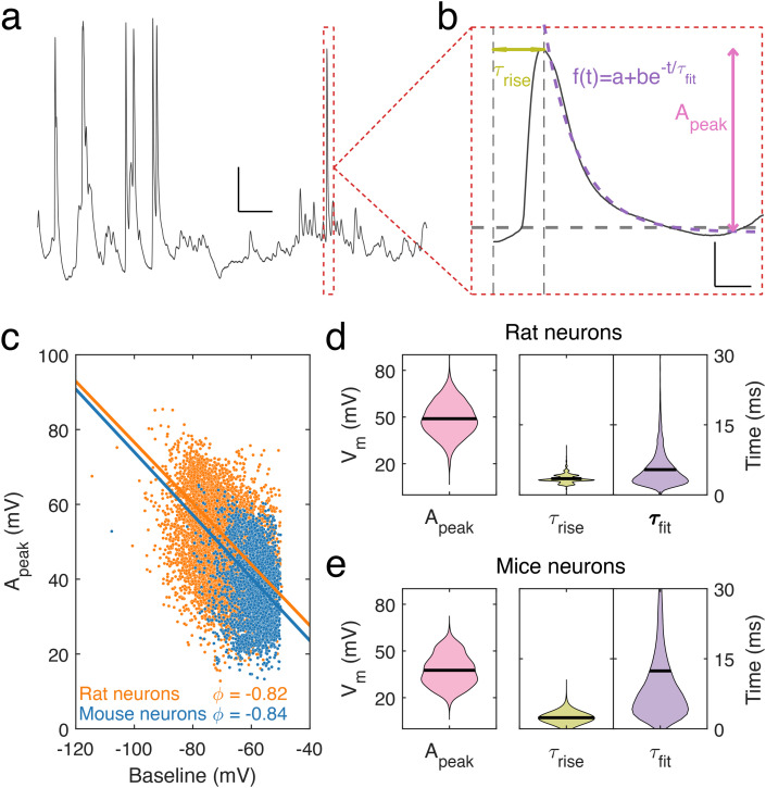
Dendrites and dendritic spines are the essential cellular compartments in neuronal communication, conveying information through transient voltage signals. Our understanding of these compartmentalized voltage dynamics in fine, distal neuronal dendrites remains poor due to the difficulties inherent to accessing and stably recording from such small, nanoscale cellular compartments for a sustained time. To overcome these challenges, we use nanopipettes that permit long and stable recordings directly from fine neuronal dendrites. We reveal a diversity of voltage dynamics present locally in dendrites, such as spontaneous voltage transients, bursting events and oscillating periods of silence and firing activity, all of which we characterized using segmentation analysis. Remarkably, we find that neuronal dendrites can display spontaneous hyperpolarisation events, and sustain transient hyperpolarised states. The voltage patterns were activity-dependent, with a stronger dependency on synaptic activity than on action potentials. Long-time recordings of fine dendritic protrusions show complex voltage dynamics that may represent a previously unexplored contribution to dendritic computations.


Conflict of interest statement

There are no conflicts to declare.

Figures

Fig. 1
Fig. 1. Electrophysiology of dendrites using nanopipettes. (a) Bright-field microscopy image of a nanopipette recording from a cultured (23 days in vitro) primary mouse neuronal dendrite. Scale bar, 5 μm. (b) Current–voltage relation for a typical nanopipette. Resistance is measured using a linear fit and this is used to determine the size of each nanopipette. (c) Distribution of nanopipette tip diameters, mean size is 17.1 ± 8.8 nm, n = 131. Each point within the violin represents a distinct recorded diameter. (d) Recording from a neuronal dendrite taken with a nanopipette, with a duration of almost one hour. Red dashed box shows a 2 s trace with transient firing activity. Baseline −80 mV, scale bar, 20 mV, 100 s, inset, scale bar: 10 mV, 100 ms. (e) Distribution of the resting membrane potential (RMP) in dendrites recorded with nanopipettes. Mean RMP is −74.5 ± 17.3 mV, n = 54. Data points within the violin are the RMP from each independent recording. (f) Measured potential during approach of the nanopipette to a dendrite (orange box) and following contact with cell (blue box), marked by sharp decrease in potential. Scale bar: 20 mV, 50 s. (g) Power spectral density (PSD) of measured potential in the bath before contacting dendrite (orange PSD corresponds to orange box in f) and after forming a patch (blue PSD corresponds to blue box in f). Exponential fitting to PSDs identifies a change in the decay of the frequency contributions to the signal.
Fig. 2
Fig. 2. Dendritic spontaneous activity. (a) Sample of a typical nanopipette electrophysiological recording from a neuronal dendrite displaying spontaneous activity. Scale bar: 10 mV, 100 ms. (b) The characteristics of a single event (red dashed box from a) decomposed as a combination of a rising and decaying segment. Three parameters characterize the event, the rise time, τrise, the peak amplitude, Apeak, of the event and the decay time, τfit, estimated by fitting an exponential function. Scale bar: 10 mV, 2.5 ms. (c) Distribution of determined Apeak values as a function of their baselines in mice (blue) and rat (orange) neuronal dendrites. Lines show the principal axes of each distribution calculated from their respective inertia tensors. (d) Distribution of Apeak, τrise and τfit from rat cortical neuron dendrites (mean values given by black lines). Mean Apeak = 49 ± 11 mV, τrise = 3.5 ± 0.9 ms, τfit = 5.4 ± 3.7 ms (n = 7537). (e) Same as in (d) but for mouse cortical neuron dendrites. Mean Apeak = 37 ± 10 mV, τrise = 2.4 ± 0.6 ms and τfit = 12.4 ± 14.8 ms (n = 2882).
Fig. 3
Fig. 3. Dendritic fast bursting events. (a) Example of repetitive fast oscillating burst recorded from a neuronal dendrite. RMP −74 mV, scale bar: 10 mV, 50 ms. (b) Single burst from red box (a) characterized by a large bursting amplitude, Aburst, a total bursting duration, Tburst, and an inter-event interval, ΔTBurst, within the burst. Scale bar: 10 mV, 10 ms. (c)–(e) Distribution of the three parameters Aburst, Tburst, and ΔTBurst. Mean values of each with standard deviation are: Aburst: 36.9 ± 16.7 mV, Tburst = 70.3 ± 20 ms and ΔTBurst = 9.2 ± 3.8 ms (n = 445). (f) ABurst, as a function of the baseline voltage for each burst. Inset: example of a bursting event, with baseline and ABurst labelled. (g) Distribution of the number of peaks per burst: NPeaks = 7.5 ± 3.1.
Fig. 4
Fig. 4. Two spontaneous regimes: silent and active regimes. (a) Examples of silent and stable behaviour, and (b) high levels of activity in traces. n = 18 969 (rat) and n = 3536 (mice) silent epochs. n = 2702 (rat) and n = 195 (mice) active periods. Mean duration of silent regime in rats is 31 ± 1 ms and 63 ± 12 ms in mice. Mean duration of active regime is 121 ± 3 ms in rats and is 529 ± 78 ms in mice. (c) Normalized counts of the occurrence of the stable regime being observed at a given baseline voltage in rat neuronal dendrites. (d) Normalized counts of the active regime being observed at a given baseline voltage in rat neuronal dendrites. (e) Normalized counts of the stable regime being recorded at a given baseline voltage in mouse neuronal dendrites. (f) Normalized counts of the active regime being recorded at a given baseline voltage in mouse neuronal dendrites. Mean frequency (in events per minute) of silent regime observed in rats is 96.5 ± 51.1 and in mice is 69.4 ± 32.9. Mean frequency of active regime observed in rats is 13.8 ± 6.6 and in mice is 5.4 ± 1.2.
Fig. 5
Fig. 5. Hyperpolarised events in neuronal dendrites. (a) Spontaneous hyperpolarisation event recorded from a dendrite. The hyperpolarisation occurred in an otherwise silent period with a baseline at −80 mV (horizontal dash line) and became hyperpolarised at a voltage below −100 mV. Scale bar: 10 mV, 100 ms. (b) Sustained period of hyperpolarisation lasting seconds. Trace shows alternating states of hyperpolarisation and spontaneous activity. Scale bar: 20 mV, 20 s. (c) Traces of hyperpolarisations (light orange) and their average (orange bold curve). (d) Sustained hyperpolarisation events (light blue), and their average (Dark blue, bold curve). (e) Full width at half minimum of hyperpolarisations against the baseline at each event. (f) Amplitude distribution of sustained hyperpolarisation events (taken as the most negative value recorded during the event) versus surrounding baseline.
Fig. 6
Fig. 6. Dendrite spontaneous activity is dependent on action potentials and synaptic activity. (a) Sample trace from a mouse neuronal dendrite in basal condition (RMP = −63 mV). (b) Reduced activity in the presence of TTX (1 μM) (RMP = −62 mV). (c) Spontaneous activity is inhibited by synaptic blockers (CPP, 10 μM, NBQX, 10 μM and picrotoxin, 100 μM, (RMP = −72 mV). Scale bar: 10 mV, 50 ms. (d) Level of spontaneous activity in each condition quantified by events count in 10 s windowed segments (basal condition n = 167 bins, TTX n = 74 bins, synaptic blocker n = 345 bins). Means indicated by black horizontal lines. Binned event count means are 30 ± 5 (basal), 11 ± 4 (TTX), 0 ± 0 (synaptic blockers). Kruskal–Wallis test performed followed by Dunn's post hoc test (Basal vs. TTX and basal vs. synaptic blockers, p < 0.0001; TTX vs. synaptic blockers, p = 0.0003). (e) Amplitude of events recorded from neuronal dendrites in basal (mean: 18.7 ± 0.1 mV, n = 5045) and TTX (mean: 18.4 ± 0.2 mV, n = 8010) conditions (p = 0.9812, Mann–Whitney test) (f) rise time of events observed in basal (mean: 7.5 ± 0.1 ms, n = 5045) and TTX (7.2 ± 0.2 ms, n = 8010) conditions (p = 0.1076, Mann–Whitney test). (g) Decay time determined for events recorded in basal (mean: 6.6 ± 0.1 ms, n = 5045) and TTX (mean: 7.6 ± 0.3 ms, n = 8010) conditions (p < 0.0001, Mann–Whitney test).

References

    1. Chua J. J. E. Kindler S. Boyken J. Jahn R. The architecture of an excitatory synapse. J. Cell Sci. 2010;123:819–823. doi: 10.1242/jcs.052696. - DOI - PubMed
    1. Söhl G. Maxeiner S. Willecke K. Expression and functions of neuronal gap junctions. Nat. Rev. Neurosci. 2005;6:191–200. doi: 10.1038/nrn1627. - DOI - PubMed
    1. Neher E. Sakmann B. Single-channel currents recorded from membrane of denervated frog muscle fibres. Nature. 1976;260:799–802. doi: 10.1038/260799a0. - DOI - PubMed
    1. Yuste R., Dendritic Spines, The MIT Press, 2010, 10.7551/mitpress/9780262013505.001.0001 - DOI
    1. Segev I. What do dendrites and their synapses tell the neuron? J. Neurophysiol. 2006;95:1295–1297. doi: 10.1152/classicessays.00039.2005. - DOI - PubMed