

Case Report: Simultaneous penetrating keratoplasty with autologous simple limbal epithelial transplantation as an alternative to keratoprosthesis
- PMID: 37455854
- PMCID: PMC10349272
- DOI: 10.12688/f1000research.133637.3
Case Report: Simultaneous penetrating keratoplasty with autologous simple limbal epithelial transplantation as an alternative to keratoprosthesis
Abstract
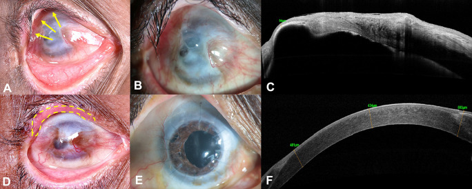
Introduction and importance: This case report highlights the multidisciplinary approach required to achieve successful anatomical and functional outcomes, in an eye with total limbal stem cell deficiency (LSCD) associated with underlying corneal scarring and thinning. Presentation of case: A 59-year-old gentleman had poor visual recovery in the right eye (RE) following accidental carbide blast, 1-year before presenting to us. The visual acuity was counting fingers and clinical examination revealed cicatricial entropion involving the upper eyelid, total LSCD, corneal scarring with a central descemetocele and cataract in the RE. Prior to ocular surface reconstruction, entropion correction was performed. Three months later, penetrating keratoplasty combined with cataract surgery and intraocular lens implantation (penetrating keratoplasty (PK) triple), with autologous simple limbal epithelial transplantation (SLET) was performed. The visual acuity was 20/100, 18 months after the surgery, with a clear well-epithelized corneal graft and stable ocular surface. Discussion: LSCD is caused by a decrease in the population and /or function of the limbal epithelial stem cells. Limbal stem cell transplantation (LSCT) is warranted in eyes with total LSCD. In eyes with coexisting corneal scarring, LSCT alone may be inadequate to restore the vision. These eyes require simultaneous or sequential lamellar or full-thickness corneal transplantation for visual rehabilitation. Though, the existing literature favors a sequential approach, where LSCT is performed first followed by corneal transplantation, under certain circumstances such as a thin underlying cornea like in our case, corneal transplantation may have to be combined with LSCT to achieve optimal outcomes. Conclusion: Combining autologous SLET with PK can be performed for visual rehabilitation in eyes with unilateral total LSCD and underlying corneal thinning. Corneal and limbal graft survival is prolonged if existing adnexal comorbidities are addressed before any surgical intervention is planned and adequate time interval is allowed for the surface inflammation to subside.
Keywords: cicatricial entropion; descemetocele; limbal stem cell deficiency (LSCD); limbal stem cell transplantation (LSCT); ocular burn; penetrating keratoplasty.
Copyright: © 2023 Sharma S et al.
Conflict of interest statement
No competing interests were disclosed.
Figures
Similar articles
-
Corneal stromal changes following simple limbal epithelial transplantation on Scheimpflug densitometry: Early results.Indian J Ophthalmol. 2025 Jan 1;73(1):77-82. doi: 10.4103/IJO.IJO_105_24. Epub 2024 Aug 14. Indian J Ophthalmol. 2025. PMID: 39186626 Free PMC article.
-
Outcomes of surgical interventions for the treatment of limbal stem cell deficiency.Indian J Med Res. 2021 Jul;154(1):51-61. doi: 10.4103/ijmr.IJMR_1139_18. Indian J Med Res. 2021. PMID: 34782530 Free PMC article.
-
Autologous Glueless Simple Limbal Epithelial Transplantation for Unilateral Stem Cell Deficiency Using Femtosecond Laser-Assisted Limbal Stem Cell Harvesting: The Report of the First 3 Clinical Cases.Cornea. 2025 Feb 21;44(8):1058-1069. doi: 10.1097/ICO.0000000000003838. Cornea. 2025. PMID: 40591740
-
Limbal stem cell transplantation: an evidence-based analysis.Ont Health Technol Assess Ser. 2008;8(7):1-58. Epub 2008 Oct 1. Ont Health Technol Assess Ser. 2008. PMID: 23074512 Free PMC article.
-
A systematic literature review of surgical interventions for limbal stem cell deficiency in humans.Am J Ophthalmol. 2008 Aug;146(2):251-259. doi: 10.1016/j.ajo.2008.03.018. Epub 2008 May 16. Am J Ophthalmol. 2008. PMID: 18486098
Cited by
-
Sequelae of carbide-related thermo-chemical injury: A retrospective analysis.Indian J Ophthalmol. 2023 Sep;71(9):3192-3197. doi: 10.4103/IJO.IJO_57_23. Indian J Ophthalmol. 2023. PMID: 37602607 Free PMC article.
-
Pathophysiologically relevant bisphenol S exposure accelerates aging by disrupting brown adipose tissue-regulated energy metabolism.Proc Natl Acad Sci U S A. 2025 Jun 10;122(23):e2420437122. doi: 10.1073/pnas.2420437122. Epub 2025 Jun 2. Proc Natl Acad Sci U S A. 2025. PMID: 40455996
-
Limbal stem cell deficiency approaches and limbal niche restoration.Indian J Ophthalmol. 2025 Apr 1;73(4):468-482. doi: 10.4103/IJO.IJO_464_25. Epub 2025 Mar 27. Indian J Ophthalmol. 2025. PMID: 40146135 Free PMC article. Review.
References
Publication types
MeSH terms
LinkOut - more resources
Full Text Sources
Medical
Research Materials