Clinical evaluation of white matter lesions on 3D inversion recovery ultrashort echo time MRI in multiple sclerosis
- PMID: 37456321
- PMCID: PMC10347363
- DOI: 10.21037/qims-22-1317
Clinical evaluation of white matter lesions on 3D inversion recovery ultrashort echo time MRI in multiple sclerosis
Abstract
Background: We clinically evaluated the quality of white matter lesions (WML) of the cerebrum on 3D inversion recovery ultrashort echo time (IR-UTE) magnetic resonance imaging (MRI) in multiple sclerosis (MS) patients.
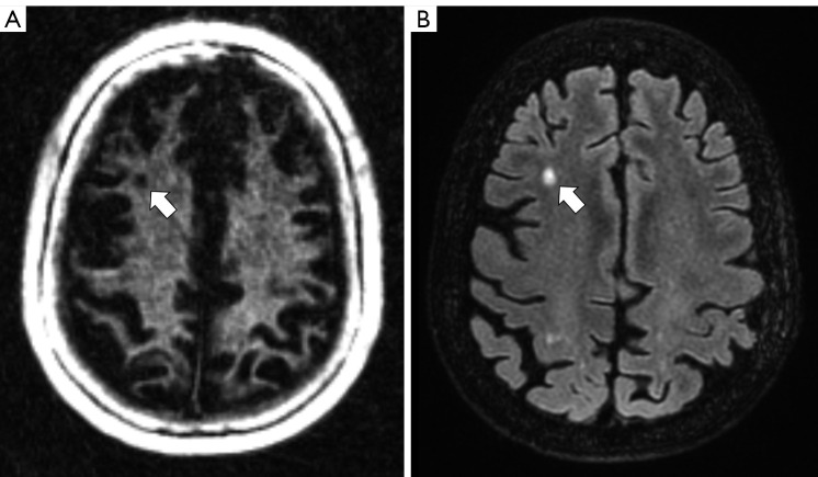
Methods: Forty-nine patients with MS were included in this study. A 3T MRI scanner was used. Two radiologists (readers) evaluated the quality of WML on IR-UTE images using a three-point Likert scale (1-good quality, 2-moderate quality, 3-insufficient quality). They also rated other WML-related factors potentially influencing WML quality using another three-point Likert scale (1-no/minor impact, 2-moderate impact, 3-high impact). Another reader rated the presence of WML on IR-UTE to evaluate the diagnostic value (right/false positive and false negative) of IR-UTE in detecting WML. Signal intensity ratios (SIRs) derived from WML signal intensities and WML sizes were also determined and analyzed.
Results: Two hundred and seventy-five MS lesions were evaluated. 87% of the lesions were rated Likert 1 on IR-UTE (P<0.01). WML rated Likert 2 and 3 presented near the grey matter (GM) in 58% of the cases (n=21), with 14 lesions being ≤2 mm (P=0.03). 62.5% of the WML rated Likert 2/3 were in the temporal lobe (P=0.02). The mean SIR of WML on IR-UTE was 1.14±0.22, while the mean SIR on fluid-attenuated inversion recovery (FLAIR) was 6.97±1.88. There was no significant correlation of SIRs between IR-UTE and FLAIR (R=0.14, P=0.245). 92.4% of the WML were correctly detected on IR-UTE (n=254). 19 out of the 21 false positive/negative rated WML were located near the GM or in the temporal lobe. WML presented 7.7% smaller in mean on IR-UTE compared to FLAIR. Factors affecting WML quality with a moderate or high impact (Likert 2 and 3) were not found.
Conclusions: Most WML are clearly detectable on IR-UTE sequences. The main limitations are WML in the temporal lobe and near the GM.
Keywords: Multiple sclerosis; brain; diagnostics; magnetic resonance imaging (MRI); white matter.
2023 Quantitative Imaging in Medicine and Surgery. All rights reserved.
Conflict of interest statement
Conflicts of Interest: All authors have completed the ICMJE uniform disclosure form (available at https://qims.amegroups.com/article/view/10.21037/qims-22-1317/coif). JD serves as an unpaid editorial board member of Quantitative Imaging in Medicine and Surgery. The other authors have no conflicts of interest to declare.
Figures