Case Reports
. 2023 Jun 29:14:878446.
doi: 10.3389/fneur.2023.878446. eCollection 2023.

Uterus infantilis: a novel phenotype associated with AARS2 new genetic variants. A case report

Affiliations
Case Reports

Uterus infantilis: a novel phenotype associated with AARS2 new genetic variants. A case report

Ekaterina Kazakova et al. Front Neurol. .

Abstract

Objectives: To report the first Mexican case with two novel AARS2 mutations causing primary ovarian failure, uterus infantilis, and early-onset dementia secondary to leukoencephalopathy.

Methods: Detailed clinical, clinimetric, neuroimaging features, muscle biopsy with biochemical assays of the main oxidative phosphorylation complexes activities, and molecular studies were performed on samples from a Mexican female.

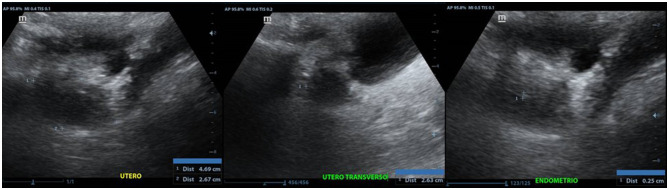
Results: We present a 41-year-old female patient with learning difficulties since childhood and primary amenorrhea who developed severe cognitive, motor, and behavioral impairment in early adulthood. Neuroimaging studies revealed frontal leukoencephalopathy with hypometabolism at the fronto-cerebellar cortex and caudate nucleus. Uterus infantilis was detected on ultrasound study. Clinical exome sequencing identified two novel variants, NM_020745:c.2864G>A (p.W955*) and NM_020745:c.1036C>A (p.P346T, p.P346Wfs*18), in AARS2. Histopathological and biochemical studies on muscle biopsy revealed mitochondrial disorder with cytochrome C oxidase (COX) deficiency.

Conclusions: Several adult-onset cases of leukoencephalopathy and ovarian failure associated with AARS2 variants have been reported. To our best knowledge, none of them showed uterus infantilis. Here we enlarge the genetic and phenotypic spectrum of AARS2-related dementia with leukoencephalopathy and ovarian failure and contribute with detailed clinical, clinometric, neuroimaging, and molecular studies to disease and novel molecular variants characterization.

Keywords: AARS2; AARS2 leukoencephalopathy; adult-onset leukodystrophy; alanyl-transfer RNA synthetase 2 mutation-related leukodystrophy; early-onset dementia; mitochondrial aminoacyl-tRNA synthetase; progressive leukoencephalopathy with ovarian failure; uterus infantilis.


Conflict of interest statement

The authors declare that the research was conducted in the absence of any commercial or financial relationships that could be construed as a potential conflict of interest.

Figures

Figure 1
Figure 1
(A) Brain magnetic resonance imaging (MRI) shows abnormal signals in the bilateral periventricular anterior white matter and the anterior part of the corpus callosum. (B) Axial T2-weighted MRI shows abnormal signals in the frontal white matter and in the pyramidal tract at the internal capsule. There was no clear pattern of cortical atrophy, and the hippocampal volume was normal. (C) The diffusion-weighted image (DWI) shows patchy areas of restricted diffusion in the abnormal white matter. (D) Visuospatial ability performance in the Minimental Status Examination (MMSE) test showing visuospatial disintegration (no intersection, loss of some sides and angles); (E) Clock Drawing Test (CDT) with perseverative drawing; (F) Writing a sentence in the MMSE test showing dysgraphia.
Figure 2
Figure 2
Anteverted uterus, with regular and poorly defined contours, with approximate dimensions of 4.6 x 2.6 x 2.6 cm in its longitudinal, transverse and anteroposterior axes, respectively.
Figure 3
Figure 3
(A) Sanger sequencing of the AARS2 in gDNA from the control, mother, and patient confirming the presence of both variants: NM_020745:c.1036C>A (p.P346T) and NM_020745:c.2864G>A (p.W955*) in the patient. The p. c.2864G>A * was identified in the mother. (B) Schematic representation of a wild type, and the alternative and constitutive splicing isoforms caused by the c.1036C>A variant. (C) Sanger sequencing showing the pathogenic alternative splicing caused by the c.1036C>A variant. Three PCR products were sequenced from a unique PCR band amplifying the AARS2 transcript. The corresponding sequences are shown in the AARS2 affected” section of the figure: the first corresponds to the allele with the c.2864G>A, p.W955* variant that appears normal in the shown area. The second read showed a deletion of seven exon nucleotides that remove the c.1036C>A and causes a change in the reading frame with a premature stop codon as a predicted consequence (p.P346Wfs*18). The third read corresponds to the c.1036C>A constitutively spliced variant translating as a P346T aminoacid substitution without a change in the reading frame.
Figure 4
Figure 4
Amino acid alignment of the AARS2 homologous proteins. The amino acid p.P346 is not conserved; only Ptroglodytes and M. Mulatta have proline in this position.
Figure 5
Figure 5
Muscle biopsy images. (A) Hematoxylin-Eosin Stain (H-E). Panoramic cross-sectional view of muscle with myopathic and angulated fascicles (marked with white arrow), difference in shape and size of the fiber, intra- and inter-fascicular connective tissue. In (B), close-up of image (A) with the presence of the hypertrophic fiber (white asterisk), surrounded by angulated fibers (black asterisk). In (C, D) images (Gomori's Modified Trichrome Stain (T-G)) the same findings are observed as in (A). In (E) panoramic view with the technique of Reduced Nicotinamide Adenine Dinucleotide-Tetrazolium Reductase (NADH-TR) with type I fibers of darker color and type II fibers of lighter color. In (F) close-up of the previous image, where a hypertrophic fiber is observed, surrounded by some angulated fibers. In (G), view of a transverse section with cytochrome C oxidase (COX) showing pale (thin black arrow) and ghost (thick black arrow) fibers. In (H) Cytochrome C Oxidase/Succinate Dehydrogenase (COX/SDH) double-labeling method we observe two blue fibers.

References

    1. Euro L, Konovalova S, Asin-Cayuela J, Tulinius M, Griffin H, Horvath R, et al. Structural modeling of tissue-specific mitochondrial alanyl-tRNA synthetase (AARS2) defects predicts differential effects on aminoacylation. Front Genet. (2015) 6:21. 10.3389/fgene.2015.00021 - DOI - PMC - PubMed
    1. Fuchs SA, Schene IF, Kok G, Jansen JM, Nikkels PGJ, van Gassen KLI, et al. Aminoacyl-tRNA synthetase deficiencies in search of common themes. Genet Med. (2019) 21:319–30. 10.1038/s41436-018-0048-y - DOI - PMC - PubMed
    1. Konovalova S, Tyynismaa H. Mitochondrial aminoacyl- tRNA synthetases in human disease. Mol Genet Metab. (2013) 108:206. 10.1016/j.ymgme.2013.01.010 - DOI - PubMed
    1. Götz A, Tyynismaa H, Euro L, Ellonen P, Hyötyläinen T, Ojala T, et al. Exome sequencing identifies mitochondrial alanyl-tRNA synthetase mutations in infantile mitochondrial cardiomyopathy. Am J Hum Genet. (2011) 88:635–42. 10.1016/j.ajhg.2011.04.006 - DOI - PMC - PubMed
    1. Dallabona C, Diodato D, Kevelam SH, Haack TB, Wong LJ, Salomons GS, et al. Novel (ovario) leukodystrophy related to AARS2 mutations. Neurology. (2014) 82:2063–71. 10.1212/WNL.0000000000000497 - DOI - PMC - PubMed

Publication types

LinkOut - more resources