. 2023 Jun 18;26(7):107168.
doi: 10.1016/j.isci.2023.107168. eCollection 2023 Jul 21.

Wet adhesive hydrogels to correct malacic trachea (tracheomalacia) A proof of concept

Affiliations

Wet adhesive hydrogels to correct malacic trachea (tracheomalacia) A proof of concept

Ece Uslu et al. iScience. .

Abstract

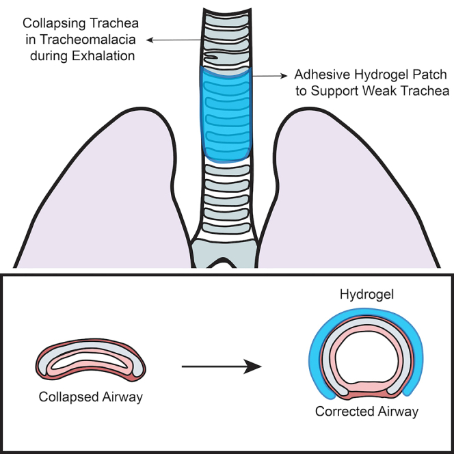
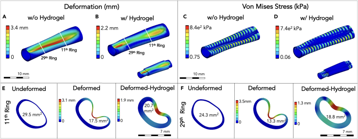
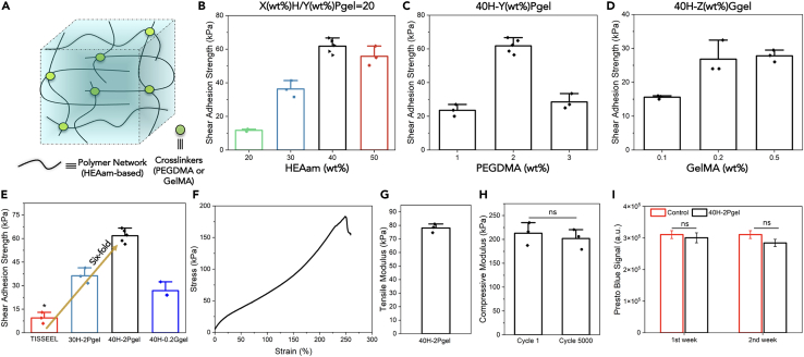
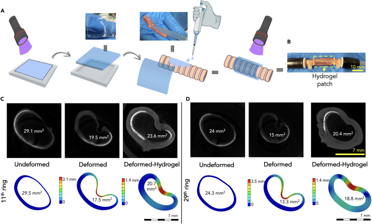
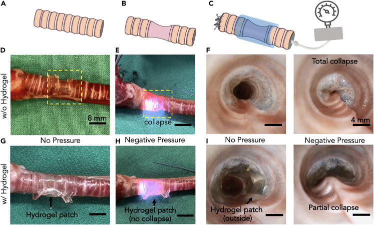
Tracheomalacia (TM) is a condition characterized by a weak tracheal cartilage and/or muscle, resulting in excessive collapse of the airway in the newborns. Current treatments including tracheal reconstruction, tracheoplasty, endo- and extra-luminal stents have limitations. To address these limitations, this work proposes a new strategy by wrapping an adhesive hydrogel patch around a malacic trachea. Through a numerical model, first it was demonstrated that a hydrogel patch with sufficient mechanical and adhesion strength can preserve the trachea's physiological shape. Accordingly, a new hydrogel providing robust adhesion on wet tracheal surfaces was synthesized employing the hydroxyethyl acrylamide (HEAam) and polyethylene glycol methacrylate (PEGDMA) as main polymer network and crosslinker, respectively. Ex vivo experiments revealed that the adhesive hydrogel patches can restrain the collapsing of malacic trachea under negative pressure. This study may open the possibility of using an adhesive hydrogel as a new approach in the difficult clinical situation of tracheomalacia.

Keywords: Applied sciences; Biomaterials.


Conflict of interest statement

The authors declare no competing interests.

Figures

None
Graphical abstract
Figure 1
Figure 1
Representation of a normal and a malacic trachea during the positive and negative pressure phases of the respiratory cycle
Figure 2
Figure 2
Experimental set-up and steps to obtain a simple trachea geometry for numerical model (A) Micro-CT (μ-CT) machine, a set-up to fix the rabbit trachea inside the machine and to create the negative pressure using a syringe filled with air. (B) Five equidistant cross-sections obtained from the μ-CT scanner. (C) 3D reconstruction of the cartilage-to-tissue volume ratio. (D) Generation of the final realistic model of equivalent geometrical properties.
Figure 3
Figure 3
Indicative results of the total deformation and the stress distribution of a trachea at −15 mmHg (A) Deformation of a trachea without hydrogel patch under applied pressure. (B) Deformation of a trachea with hydrogel patch under applied pressure. (C) Von Mises stress distribution of a trachea without hydrogel patch under applied pressure. (D) Von Mises stress distribution of a trachea with hydrogel patch under applied pressure. (E) Cross-sections of the rabbit trachea model at the 11th ring. (F) Cross-sections of the rabbit trachea model at the 29th tracheal rings.
Figure 4
Figure 4
Characterization of the adhesive hydrogel (A) Schematic of the adhesive hydrogel. (B) Screening to find out the hydrogel formulation having highest shear adhesion strength on the wet rabbit trachea surface by changing HEAam concentration at fixed cross-linking density (HEAam(%)/PEGDM(%) = 20). (C) Screening to find out the hydrogel formulation having highest shear adhesion strength on the wet rabbit trachea surface by changing PEGDMA concentration at constant HEAam concentration (40 wt %). (D) Screening to find out the hydrogel formulation having highest shear adhesion strength on the wet rabbit trachea surface by changing concentration of another crosslinker GelMA at constant HEAam concentration (40 wt %). (E) Shear adhesion strength of three hydrogels on the trachea and comparison with a commercial glue, TISSEEL. (F) Typical stress-strain curve of 40H-2Pgel hydrogel. (G) Tensile modulus of 40H-2Pgel hydrogel. (H) Compressive modulus of 40H-2Pgel after cycle 1 and 5000. (I) Cytotoxicity analysis of 40H-2Pgel after two weeks of incubation. ∗p < 0.05 compared to other samples. Data are represented as mean ± SEM.
Figure 5
Figure 5
The potential of the adhesive hydrogel to prevent collapse in mild malacic conditions (A) Schematic of a hydrogel-patch (15 × 25 × 0.7 mm3) synthesis and preparation for a hydrogel-wrapped rabbit trachea, notably photo-polymerization was performed twice. For μ-CT analysis, rabbit trachea was kept in collagenase type 1 enzyme for 30 h to mimic mild TM conditions. (B) A photo of a hydrogel patch-wrapped trachea where both openings were fixed using plastic clips to create negative pressure. (C) μ-CT scans and numerical model of the 11th rings of trachea with and without application of the adhesive hydrogel at −15 mmHg. (D) μ-CT scans and numerical model of the 29th rings of trachea with and without application of the adhesive hydrogel at −15 mmHg.
Figure 6
Figure 6
Procedure and results of ex vivo experiments (A) Schematic of a normal trachea. (B) Schematic of a malacic trachea (tracheomalacia). (C) Flexible bronchoscope with an in-built suction channel with the hydrogel patch wrapping the malacic trachea. The distal end of the trachea was closed with surgical sutures to allow maximal negative pressure effect on applying the suction. (D) An external view of a malacic trachea, cartilage rings were removed with preservation of the underlying mucosa to mimic tracheomalacia in a rabbit trachea. (E) an external image of a malacic trachea under pressure that immediately collapsed even at a very low negative pressure (−1 to −2 kPa). The bright light is of the bronchoscope that visualized the tracheal lumen (see Video S2). (F) Endoluminal image of the trachea under negative pressure captured by bronchoscope (see Video S3). Evidently, tracheal luminal structure was completely collapsed, a perfect example of the induced tracheomalacia. (G) An external image of hydrogel patch (15 × 35 × 0.7 mm3) wrapping the malacic trachea without any pressure. (H) An external view of the hydrogel patch-wrapped malacic trachea with negative pressure. The bright light of the bronchoscope can be seen through the transparent tracheal mucosa. Airway collapse improved up to 50%, (see Video S4). (I) Endoscopic luminal images further confirmed the improvement in collapse (see Video S5). Scale bars for external view of a malacic trachea and bronchoscope images with and without hydrogel are 8 and 4 mm, respectively.

References

    1. Serrano-Casorran C., Lopez-Minguez S., Rodriguez-Zapater S., Bonastre C., Guirola J.A., De Gregorio M.A. A new airway spiral stent designed to maintain airway architecture with an atraumatic removal after full epithelization—Research of feasibility and viability in canine patients with tracheomalacia. Pediatr. Pulmonol. 2020;55:1757–1764. doi: 10.1002/ppul.24816. - DOI - PubMed
    1. Kamran A., Jennings R.W. Tracheomalacia and Tracheobronchomalacia in Pediatrics: An Overview of Evaluation, Medical Management, and Surgical Treatment. Front. Pediatr. 2019;7:512–519. doi: 10.3389/fped.2019.00512. - DOI - PMC - PubMed
    1. Choi S., Lawlor C., Rahbar R., Jennings R. Diagnosis, Classification, and Management of Pediatric Tracheobronchomalacia: A Review. JAMA Otolaryngol. Head Neck Surg. 2019;145:265–275. doi: 10.1001/jamaoto.2018.3276. - DOI - PubMed
    1. Burg G., Hossain M.M., Wood R., Hysinger E.B. Evaluation of agreement on presence and severity of tracheobronchomalacia by dynamic flexible bronchoscopy. Ann. Am. Thorac. Soc. 2021;18:1749–1752. doi: 10.1513/AnnalsATS.202009-1142RL. - DOI - PubMed
    1. Murgu S.D., Colt H.G. Tracheobronchomalacia and Excessive Dynamic Airway Collapse. Respirology. 2006;11:388–406. doi: 10.1111/j.1400-1843.2006.00862.x. - DOI - PubMed

LinkOut - more resources