Review
. 2023 Jun 29:17:1189606.
doi: 10.3389/fnins.2023.1189606. eCollection 2023.

Meningiomas with CNS invasion

Affiliations
Review

Meningiomas with CNS invasion

Konstantinos Gousias et al. Front Neurosci. .

Abstract

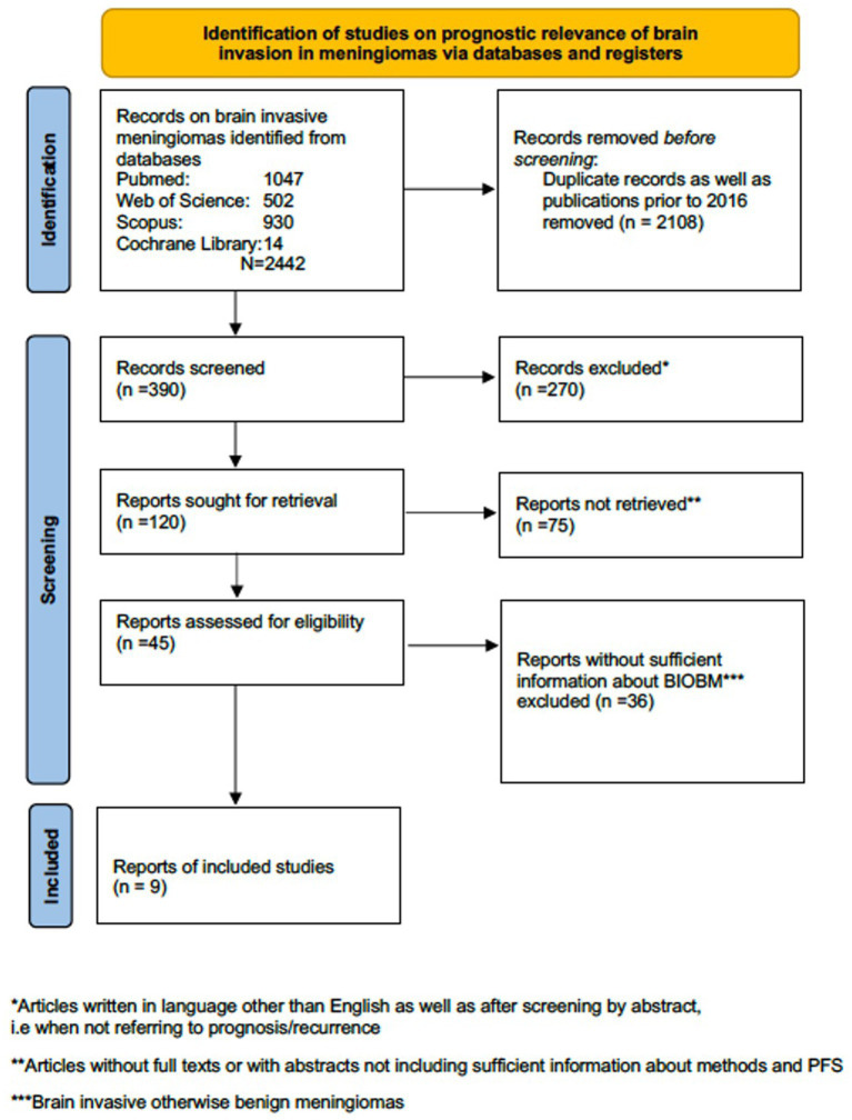
CNS invasion has been included as an independent criterion for the diagnosis of a high-grade (WHO and CNS grade 2 and 3) meningioma in the 2016 and more recently in the 2021 WHO classification. However, the prognostic role of brain invasion has recently been questioned. Also, surgical treatment for brain invasive meningiomas may pose specific challenges. We conducted a systematic review of the 2016-2022 literature on brain invasive meningiomas in Pubmed, Scopus, Web of Science and the Cochrane Library. The prognostic relevance of brain invasion as a stand-alone criterion is still unclear. Additional and larger studies using robust definitions of histological brain invasion and addressing the issue of sampling errors are clearly warranted. Although the necessity of molecular profiling in meningioma grading, prognostication and decision making in the future is obvious, specific markers for brain invasion are lacking for the time being. Advanced neuroimaging may predict CNS invasion preoperatively. The extent of resection (e.g., the Simpson grading) is an important predictor of tumor recurrence especially in higher grade meningiomas, but also - although likely to a lesser degree - in benign tumors, and therefore also in brain invasive meningiomas with and without other histological features of atypia or malignancy. Hence, surgery for brain invasive meningiomas should follow the principles of maximal but safe resections. There are some data to suggest that safety and functional outcomes in such cases may benefit from the armamentarium of surgical adjuncts commonly used for surgery of eloquent gliomas such as intraoperative monitoring, awake craniotomy, DTI tractography and further advanced intraoperative brain tumor visualization.

Keywords: CNS invasion; Simpson grade of resection; functional outcome; invasive meningioma; surgery.


Conflict of interest statement

The authors declare that the research was conducted in the absence of any commercial or financial relationships that could be construed as a potential conflict of interest.

Figures

Figure 1
Figure 1
PRISMA flow diagram regarding studies on prognostic relevance of brain invasive meningioma.
Figure 2
Figure 2
Imperfect correlations between imaging findings, histopathological atypia, and brain invasion (I). (A) 64 years old male patient with a very large left anterior clinoidal meningioma assigned to CNS grade 2 based on cytological atypia and an increased mitotic count. However, there was no brain invasion. Somewhat fittingly, MR imaging reveals cysts, a cleft sign and heterogenous contrast enhancement as well as FLAIR and T2 intratumoral heterogeneity, but there was only limited peritumoral edema. (B) 82 years old female patient with a large right>left olfactory groove meningioma CNS grade 2. The neuropathological evaluation revealed no atypia, but prominent brain invasion. There is surprisingly little edema. Contrast enhancement is somewhat heterogenous, but the tumor looks rather homogenous on the T2 and FLAIR weighted images.
Figure 3
Figure 3
Imperfect correlations between imaging findings, histopathological atypia, and brain invasion (II). (A) 74 years old female patient with a left ventricular (trigonal) CNS grade 2 meningioma. This tumor had atypical histopathological features and was found to invade the brain. Possibly in contrast, the MR showed little edema. However, the actual zone of contact between the tumor and the brain parenchyma is very small. T2, FLAIR and contrast-enhanced T1 imaging reveals little heterogeneity. (B) 64 years old female patient right parietal parasagittal meningioma CNS grade 2. There was no brain invasion, however, histopathological atypia. Note, that there is substantial edema, while the tumor tissue looks otherwise inconspicuous on T2, FLAIR and contrast-enhanced T1 weighted MR images.

Similar articles

Cited by

References

    1. Adeli A., Hess K., Mawrin C., Streckert E. M. S., Stummer W., Paulus W., et al. . (2018). Prediction of brain invasion in patients with meningiomas using preoperative magnetic resonance imaging. Oncotarget 9, 35974–35982. doi: 10.18632/oncotarget.26313, PMID: - DOI - PMC - PubMed
    1. Alvernia J. E., Dang N. D., Sindou M. P. (2011). Convexity meningiomas: study of recurrence factors with special emphasis on the cleavage plane in a series of 100 consecutive patients. J. Neurosurg. 115, 491–498. doi: 10.3171/2011.4.JNS101922, PMID: - DOI - PubMed
    1. Bailey D. D., Montgomery E. Y., Garzon-Muvdi T. (2023). Metastatic high-grade meningioma: a case report and review of risk factors for metastasis. Neurooncol. Adv. 5:vdad014. doi: 10.1093/noajnl/vdad014 - DOI - PMC - PubMed
    1. Bakhshi S. K., Jawed N., Shafiq F., Enam S. A. (2021). Awake craniotomy for resection of intracranial meningioma: first case series from a low-and middle-income country. Cureus 13:e18716. doi: 10.7759/cureus.18716, PMID: - DOI - PMC - PubMed
    1. Banan R., Abbetmeier-Basse M., Hong B., Dumitru C. A., Sahm F., Nakamura M., et al. . (2021). The prognostic significance of clinicopathological features in meningiomas: microscopic brain invasion can predict patient outcome in otherwise benign meningiomas. Neuropathol. Appl. Neurobiol. 47, 724–735. doi: 10.1111/nan.12700, PMID: - DOI - PubMed

LinkOut - more resources