

The impact of TP53 activation and apoptosis in primary hereditary microcephaly
- PMID: 37457016
- PMCID: PMC10338886
- DOI: 10.3389/fnins.2023.1220010
The impact of TP53 activation and apoptosis in primary hereditary microcephaly
Abstract
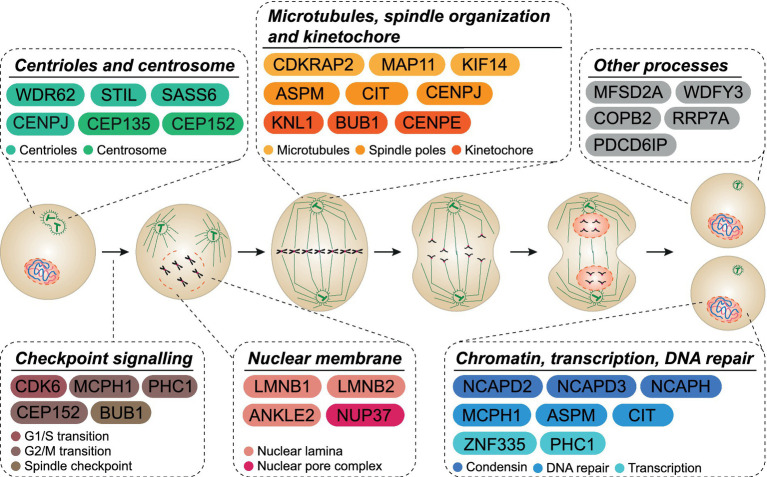
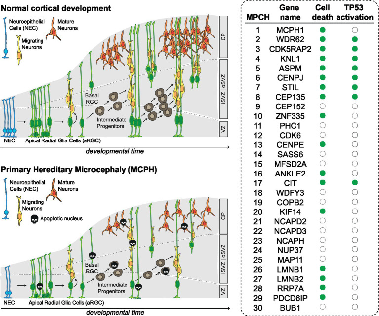
Autosomal recessive primary microcephaly (MCPH) is a constellation of disorders that share significant brain size reduction and mild to moderate intellectual disability, which may be accompanied by a large variety of more invalidating clinical signs. Extensive neural progenitor cells (NPC) proliferation and differentiation are essential to determine brain final size. Accordingly, the 30 MCPH loci mapped so far (MCPH1-MCPH30) encode for proteins involved in microtubule and spindle organization, centriole biogenesis, nuclear envelope, DNA replication and repair, underscoring that a wide variety of cellular processes is required for sustaining NPC expansion during development. Current models propose that altered balance between symmetric and asymmetric division, as well as premature differentiation, are the main mechanisms leading to MCPH. Although studies of cellular alterations in microcephaly models have constantly shown the co-existence of high DNA damage and apoptosis levels, these mechanisms are less considered as primary factors. In this review we highlight how the molecular and cellular events produced by mutation of the majority of MCPH genes may converge on apoptotic death of NPCs and neurons, via TP53 activation. We propose that these mechanisms should be more carefully considered in the alterations of the sophisticated equilibrium between proliferation, differentiation and death produced by MCPH gene mutations. In consideration of the potential druggability of cell apoptotic pathways, a better understanding of their role in MCPH may significantly facilitate the development of translational approaches.
Keywords: DNA damage; TP53; asymmetric division; cell death; microcephaly; mitosis; neural precursor; neurodevelopment.
Copyright © 2023 Iegiani, Ferraro, Pallavicini and Di Cunto.
Conflict of interest statement
The authors declare that the research was conducted in the absence of any commercial or financial relationships that could be construed as a potential conflict of interest.
Figures
References
Publication types
LinkOut - more resources
Full Text Sources
Molecular Biology Databases
Research Materials
Miscellaneous