Review
. 2023 Jul 1;16(7):dmm050005.
doi: 10.1242/dmm.050005. Epub 2023 Jul 17.

Zika virus infection histories in brain development

Affiliations
Review

Zika virus infection histories in brain development

Bruna L M Marcelino et al. Dis Model Mech. .

Abstract

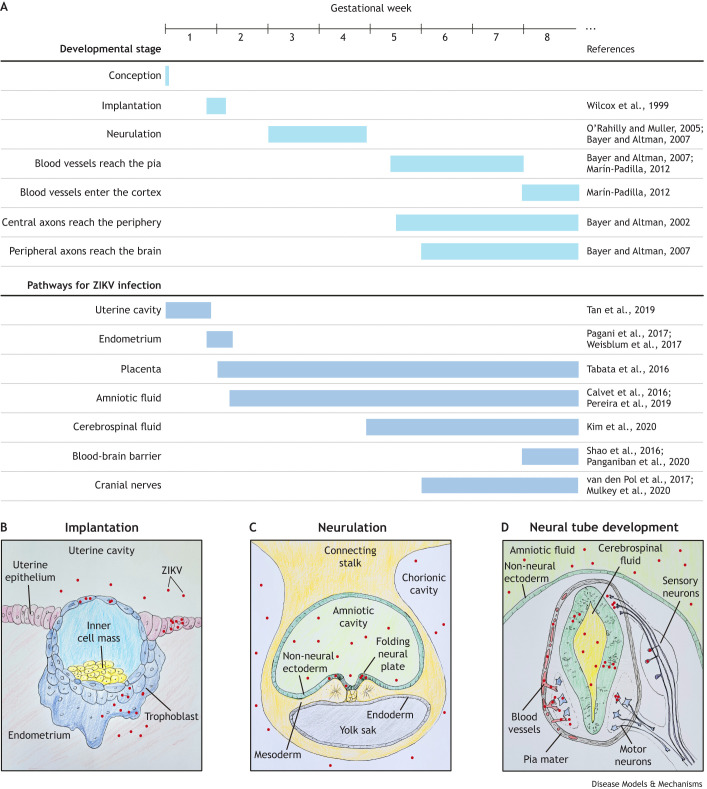
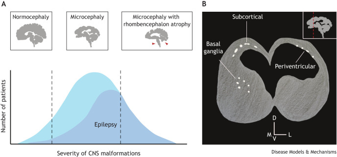
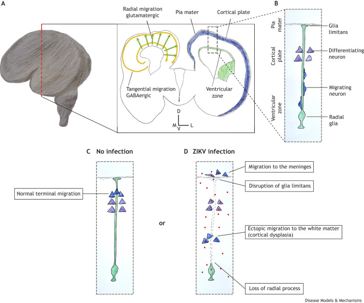
An outbreak of births of microcephalic patients in Brazil motivated multiple studies on this incident. The data left no doubt that infection by Zika virus (ZIKV) was the cause, and that this virus promotes reduction in neuron numbers and neuronal death. Analysis of patients' characteristics revealed additional aspects of the pathology alongside the decrease in neuronal number. Here, we review the data from human, molecular, cell and animal model studies attempting to build the natural history of ZIKV in the embryonic central nervous system (CNS). We discuss how identifying the timing of infection and the pathways through which ZIKV may infect and spread through the CNS can help explain the diversity of phenotypes found in congenital ZIKV syndrome (CZVS). We suggest that intraneuronal viral transport is the primary mechanism of ZIKV spread in the embryonic brain and is responsible for most cases of CZVS. According to this hypothesis, the viral transport through the blood-brain barrier and cerebrospinal fluid is responsible for more severe pathologies in which ZIKV-induced malformations occur along the entire anteroposterior CNS axis.

Keywords: Blood–brain barrier; Brainstem; Calcification; Cerebellum; Neuronal migration; Susceptibility window.


Conflict of interest statement

Competing interests The authors declare no competing or financial interests.

Figures

Fig. 1.
Fig. 1.
Human early embryogenesis milestones and the possible routes for Zika virus (ZIKV) infection and spread throughout the central nervous system (CNS). (A) Timeline of key human developmental milestones and the appearance of pathways for ZIKV transport to the embryonic CNS. Relevant references that support each milestone timing and infection pathway are provided in the right column. (B-D) Representative drawings of the development of routes for ZIKV transport in the early embryo. (B) Transverse section of the embryo implanting in the endometrium. After the blastocyst hatches from the zona pellucida, trophoblast cells come into direct contact with the uterine environment. ZIKV infects trophoblast cells and possibly reaches the embryonic cells in the inner cell mass. After the embryo implants in the endometrium, it loses its direct contact with the uterine cavity. However, ZIKV can infect endometrial cells. Therefore, ZIKV can infect trophoblast cells through the uterine cavity and through the endometrium. (C) Transverse section of the trilaminar embryonic disc surrounded by the amnion and yolk sac. Human neurulation starts at the third gestational week (GW). At this stage, neural cells are in contact with the amniotic fluid, which can also become infected. ZIKV in the amniotic fluid infects the neural plate cells but not the non-neural ectoderm. It does not display tropism for mesoderm or endoderm cells either. (D) Transverse section of the neural tube at the second month of gestation. After the non-neural ectoderm closes, the amniotic fluid is separated from the neural tube and is therefore not a possible direct infection pathway anymore. From this stage on, to infect the CNS, ZIKV has to cross through the barriers between the blood and the meninges (represented on the left side of the neural tube in red and gray, respectively), the blood and the cerebrospinal fluid, or the blood and the brain parenchyma (represented on the left side of the neural tube). Alternatively, ZIKV needs to be transported along the nerves that leave (motor neurons; blue) or enter (sensory neurons; purple) (both neuron types are represented on the right side of the neural tube) the CNS.
Fig. 2.
Fig. 2.
The diversity of congenital ZIKV syndrome (CZVS) brain phenotypes and the relationship between anatomical and physiological outcomes. (A) The severity of brain malformations in CZVS can be broadly divided into three types: (1) normocephaly with punctual malformations in the prosencephalon, (2) microcephaly with malformations restricted to the prosencephalon, and (3) microcephaly with caudal malformations (red arrowheads). The graph shows the approximate frequency of CZVS and CZVS-linked epilepsy cases across the brain malformation severity spectrum. Patients with caudal malformations have high odds of developing epilepsy, whereas normocephalic ones are less likely to be affected. (B) Schematic illustration of a coronal section of the brain of a postnatal CZVS patient as seen in a computed tomography examination. The inset in the top right corner represents a sagittal section of the brain, and the dashed red line indicates the position of the coronal section. Calcifications are represented in white. Subcortical calcifications are the most common in CZVS, but periventricular ones and calcifications in the basal ganglia also occur. D, dorsal; V, ventral; M, medial; L, lateral.
Fig. 3.
Fig. 3.
ZIKV-induced neuronal migration defects. (A) Schematic representation of a human brain at the 17th GW. The dashed red line indicates the position of the coronal section represented in the rectangle on the right. The schematic of the left hemisphere shows the two types of neuronal migration in the cortical plate: the tangential migration of inhibitory GABAergic neurons (yellow arrows) and the radial migration of excitatory glutamatergic neurons (green arrows). In the schematic of the right hemisphere, the proliferative layers are represented in green and the developing cortical plate is depicted in blue. The dashed line box indicates the localization of panels B-D. (B) At this stage of brain development (17th GW), radial glia form a physical support for the migration of newly generated neurons. The schematic shows that the neurons that previously migrated and are starting to differentiate reside closer to the basal process of the radial glia (where it attaches to the pia mater). (C) In non-infected developing brains, the migrating neurons pass through the previously formed cortical layers and detach from the radial glia closer to the pia, clustering in the cortical plate. (D) In ZIKV infection, the basal processes of radial glia can become deformed or are lost, diminishing cues for proper neuronal migration. Moreover, the loss of glia limitans (the radial glial foot in the pia) increases the ‘permeability’ of the zone between the brain parenchyma and the meninges, allowing developing neurons to ectopically migrate to the meninges. Additionally, loss of radial glia processes causes premature interruption of neuronal migration, resulting in ectopic accumulation in the white matter. D, dorsal; V, ventral; M, medial; L, lateral.

Similar articles

Cited by

References

    1. Acosta-Reyes, J., Navarro, E., Herrera, M. J., Goenaga, E., Ospina, M. L., Parra, E., Mercado, M., Chaparro, P., Beltran, M., Gunturiz, M. L.et al. ( 2017). Severe neurologic disorders in 2 fetuses with Zika virus infection, Colombia. Emerg. Infect. Dis. 23, 982- 984. 10.3201/eid2306.161702 - DOI - PMC - PubMed
    1. Aguiar, R. S., Pohl, F., Morais, G. L., Nogueira, F. C. S., Carvalho, J. B., Guida, L., Arge, L. W. P., Melo, A., Moreira, M. E. L., Cunha, D. P.et al. ( 2020). Molecular alterations in the extracellular matrix in the brains of newborns with congenital Zika syndrome. Sci. Signal 13, eaay6736. 10.1126/scisignal.aay6736 - DOI - PubMed
    1. Al-Jaberi, N., Lindsay, S., Sarma, S., Bayatti, N. and Clowry, G. J. ( 2015). The early fetal development of human neocortical GABAergic interneurons. Cereb. Cortex N. Y. NY 25, 631- 645. 10.1093/cercor/bht254 - DOI - PMC - PubMed
    1. Alves, L. V., Hazin, A. N. and Alves, J. G. B. ( 2021). Neuroimaging in children born with congenital Zika syndrome: a cohort study. J. Child Neurol. 36, 1066- 1070. 10.1177/08830738211027719 - DOI - PubMed
    1. Anderson, D., Neri, J. I. C. F., Souza, C. R. M., Valverde, J. G., De Araújo, J. M. G., Nascimento, M. D. S. B., Branco, R. C. C., Arrais, N. M. R., Lassmann, T., Blackwell, J. M.et al. ( 2021). Zika virus changes methylation of genes involved in immune response and neural development in brazilian babies born with congenital microcephaly. J. Infect. Dis. 223, 435- 440. 10.1093/infdis/jiaa383 - DOI - PubMed

Publication types