. 2023 Dec 11;214(1):26-35.
doi: 10.1093/cei/uxad084.

TAGAP activates Th17 cell differentiation by promoting RhoA and NLRP3 to accelerate rheumatoid arthritis development

Affiliations

TAGAP activates Th17 cell differentiation by promoting RhoA and NLRP3 to accelerate rheumatoid arthritis development

Hong-Gang Sun et al. Clin Exp Immunol. .

Abstract

Rheumatoid arthritis (RA) is a chronic autoimmune disorder that can give rise to joint swelling and inflammation, potentially affecting the entire body, closely linked to the state of T cells. The T-cell activation Rho GTPase activating protein (TAGAP) is associated with many autoimmune diseases including RA and is directly linked to the differentiation of Th17 cells. The present study intends to investigate the influence of TAGAP on the RA progression and its mechanism to empower new treatments for RA. A collagen-induced-arthritis (CIA) rat model was constructed, as well as the extraction of CD4+ T cells. RT-qPCR, H&E staining and safranin O/fast green staining revealed that TAGAP interference reduced TAGAP production in the ankle joint of CIA rats, and joint inflammation and swelling were alleviated, which reveals that TAGAP interference reduces synovial inflammation and cartilage erosion in the rat ankle joint. Expression of inflammatory factors (TNF-α, IL-1β, and IL-17) revealed that TAGAP interference suppressed the inflammatory response. Expression of pro-inflammatory cytokines, matrix-degrading enzymes, and anti-inflammatory cytokines at the mRNA level was detected by RT-qPCR and revealed that TAGAP interference contributed to the remission of RA. Mechanistically, TAGAP interference caused a significant decrease in the levels of RhoA and NLRP3. Assessment of Th17/Treg levels by flow cytometry revealed that TAGAP promotes Th17 cells differentiation and inhibits Treg cells differentiation in vitro and in vivo. In conclusion, TAGAP interference may decrease the differentiation of Th17 cells by suppressing the expression of RhoA and NLRP3 to slow down the RA progression.

Keywords: NLRP3 inflammasome; TAGAP; Th17 cell; gene interference; rheumatoid arthritis.


Conflict of interest statement

The authors declare that they have no known competing financial interests or personal relationships that could have appeared to influence the work reported in this paper.

Figures

Graphical Abstract
Graphical Abstract
Figure 1.
Figure 1.
TAGAP interference attenuates ankle osteoarthritis in CIA rat models. (A) TAGAP expression in serum of CIA rat model (n = 6). (B) The degree of ankle and paw swelling in each group of rats on day 30. Our scoring system was as follows: 0, no signs of erythema or swelling; 1, erythema and mild swelling confined to the tarsal or ankle joints; 2, erythema and mild swelling extending from the ankle to the tarsus; 3, erythema and moderate swelling extending from the ankle to the metatarsal joints; 4, erythema and severe swelling including the ankle, paws and digits, or ankylosis of the limb (n = 6). (C) Trends in arthritis indices of rats in each group during administration. CIA-LV-TAGAP-shRNA vs. CIA-LV-shRNA-NC, *P < 0.05, ***P < 0.001, n = 3. (D) H&E staining of the ankle joint of CIA rats (blue area indicates nucleus and cell membrane, red area indicates cytoplasm, and extracellular matrix) (×100). Safranin O/fast green staining of the ankle joint of CIA rats (red area indicates cartilage area; green area indicates bone tissue, muscle and collagen fibers) (×100). All experiments were repeated three times (n = 3). CIA: collagen-induced-arthritis, TAGAP: T-cell activation Rho GTPase activating protein
Figure 2.
Figure 2.
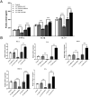
Inflammatory factor expression is affected in CIA rats under TAGAP interference. (A) The levels of TNFα, IL-1β, and IL-17 in serum and synovial tissues of rats. ***P < 0.001, n = 6. (B) The levels of TNFα, IL-6, IL-1β, MMP-3, MMP-13, and IL-10 in rat serum. ***P < 0.001, n = 3. All experiments were repeated three times. CIA: collagen-induced-arthritis, TAGAP: T-cell activation Rho GTPase activating protein
Figure 3.
Figure 3.
Decreased expression of inflammatory factors in CD4+ T cells after TAGAP interference. (A) The levels of TNFα, IL-1β, and IL-17 in the culture medium supernatant. ***P < 0.001, n = 3. (B) The levels of BCL-2, IL-6, MMP-3, MMP-13, and IFN-γ in the cells. ***P < 0.001, n = 3. All experiments were repeated three times. TAGAP: T-cell activation Rho GTPase activating protein
Figure 4.
Figure 4.
Protein content of RhoA and NLRP3 affected by TAGAP in CIA rats and CD4+ T cells. (A) Protein content of RhoA and NLRP3 in CIA rat models. ***P < 0.001, n = 3. (B) TAGAP interference or overexpression affects the protein content of RhoA and NLRP3 in CD4+ T cells. ***P < 0.001, n = 3. All experiments were repeated three times. CIA: collagen-induced-arthritis, TAGAP: T-cell activation Rho GTPase activating protein
Figure 5.
Figure 5.
Restoration of Th17 cell and Treg cell differentiation after TAGAP interference. (A) Flow cytometry detection of Th17 cell differentiation in synovial tissue. (B) Quantification of TH17 cells in synovial tissue. ***P < 0.001. (C) Flow cytometry detection of Treg cell differentiation in synovial tissue. (D) Quantification of Treg cells in synovial tissue. ***P < 0.001. (E) The presence of CD3+ T cells in the synovial tissue was visualized through immunohistochemistry staining of CD3 (×100, CD3+ T cells aggregated into clusters are represented by black arrows.). (F) Flow cytometry detection of Th17 cell differentiation in CD4+ T cells. (G) Quantification of Treg cells in synovial tissue. ***P < 0.001, n = 3. All experiments were repeated three times. TAGAP: T-cell activation Rho GTPase activating protein

References

    1. Smolen JS, Aletaha D, McInnes IB.. Rheumatoid arthritis. Lancet (London, England) 2016, 388, 2023–38. doi:10.1016/S0140-6736(16)30173-8. - DOI - PubMed
    1. Firestein GS, McInnes IB.. Immunopathogenesis of rheumatoid arthritis. Immunity 2017, 46, 183–96. doi:10.1016/j.immuni.2017.02.006. - DOI - PMC - PubMed
    1. McInnes IB, Schett G.. Cytokines in the pathogenesis of rheumatoid arthritis. Nat Rev Immunol 2007, 7, 429–42. doi:10.1038/nri2094. - DOI - PubMed
    1. Ahmad SF, Ansari MA, Nadeem A, Zoheir KMA, Bakheet SA, Al-Shabanah OA, et al. . The tyrosine kinase inhibitor tyrphostin AG126 reduces activation of inflammatory cells and increases Foxp3(+) regulatory T cells during pathogenesis of rheumatoid arthritis. Mol Immunol 2016, 78, 65–78. doi:10.1016/j.molimm.2016.08.017. - DOI - PubMed
    1. Abd-Allah AR, Ahmad SF, Alrashidi I, Abdel-Hamied HE, Zoheir KMA, Ashour AE, et al. . Involvement of histamine 4 receptor in the pathogenesis and progression of rheumatoid arthritis. Int Immunol 2014, 26, 325–40. doi:10.1093/intimm/dxt075. - DOI - PubMed

Publication types

Substances