. 2023 Jul 25;17(14):13594-13610.
doi: 10.1021/acsnano.3c02268. Epub 2023 Jul 17.

Ionizable Lipid Nanoparticles for Therapeutic Base Editing of Congenital Brain Disease

Affiliations

Ionizable Lipid Nanoparticles for Therapeutic Base Editing of Congenital Brain Disease

Rohan Palanki et al. ACS Nano. .

Abstract

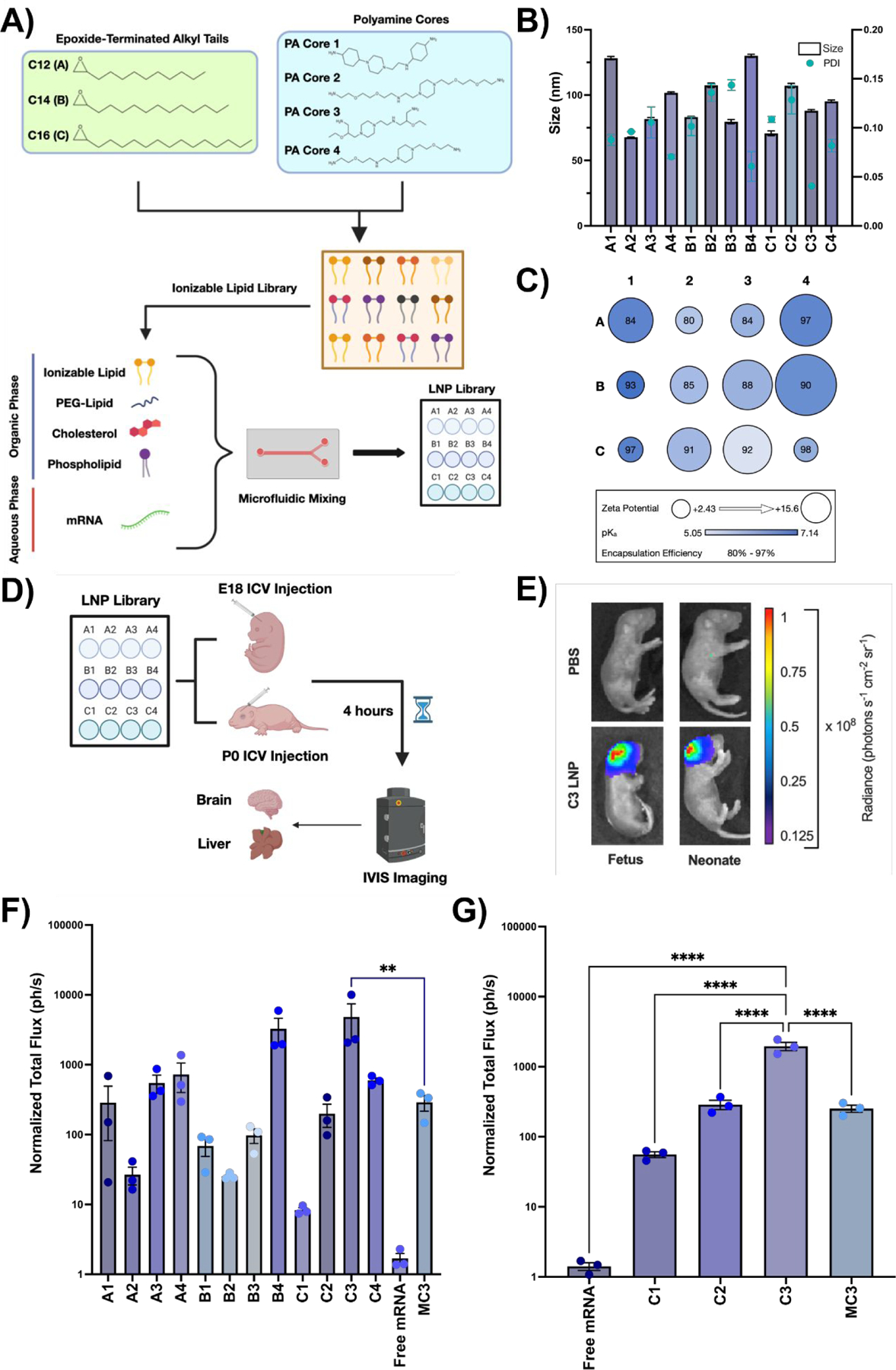
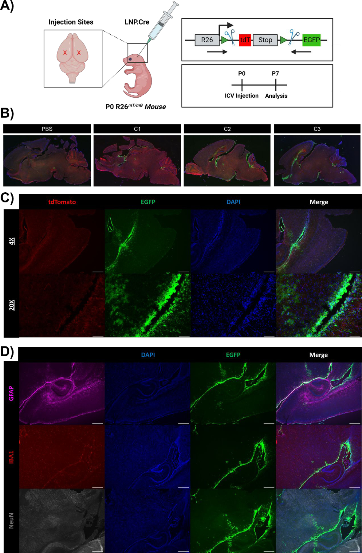
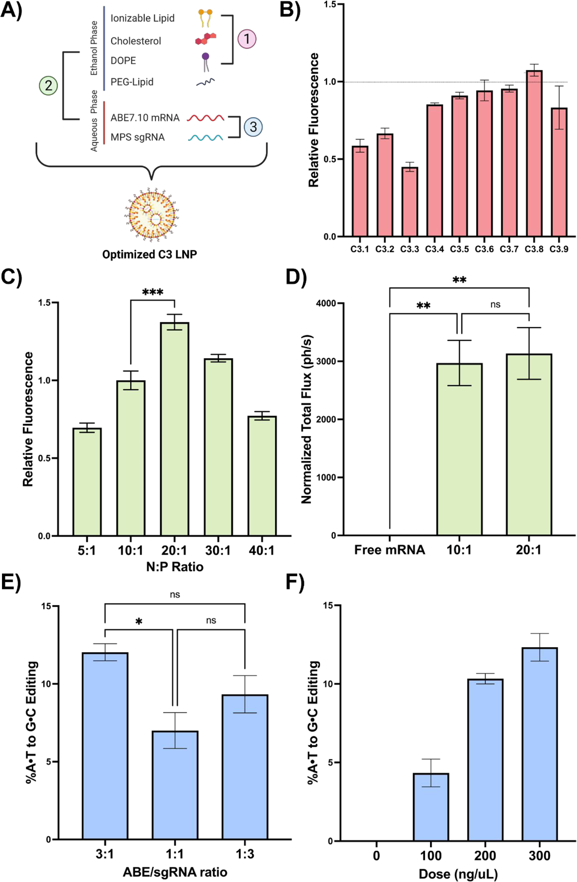
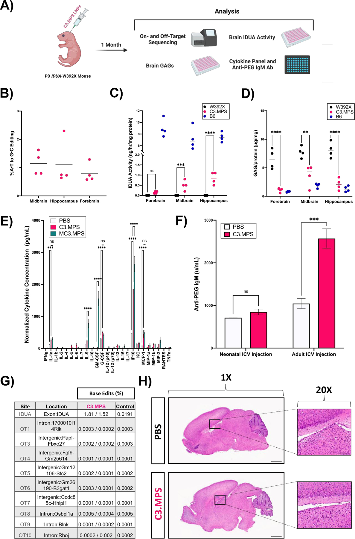
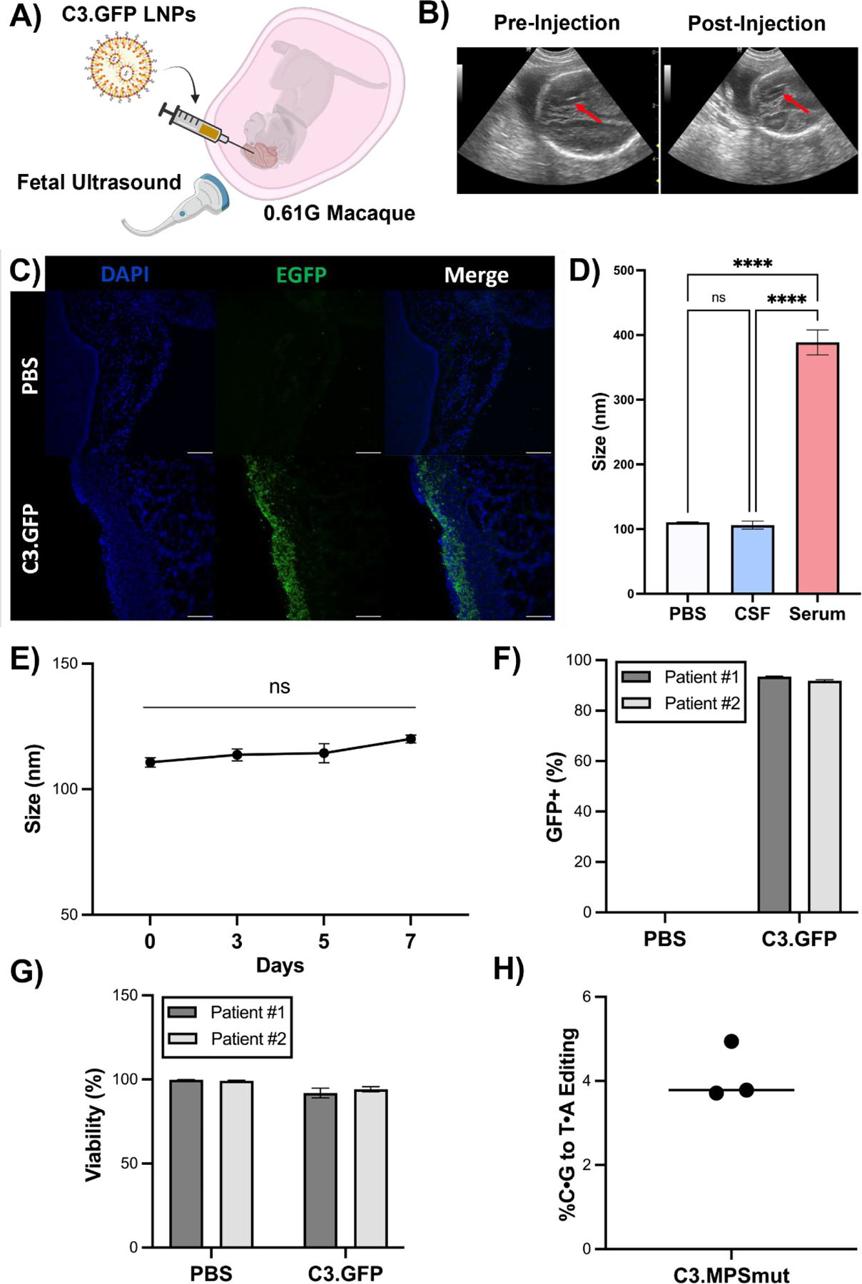
Delivery of mRNA-based therapeutics to the perinatal brain holds great potential in treating congenital brain diseases. However, nonviral delivery platforms that facilitate nucleic acid delivery in this environment have yet to be rigorously studied. Here, we screen a diverse library of ionizable lipid nanoparticles (LNPs) via intracerebroventricular (ICV) injection in both fetal and neonatal mice and identify an LNP formulation with greater functional mRNA delivery in the perinatal brain than an FDA-approved industry standard LNP. Following in vitro optimization of the top-performing LNP (C3 LNP) for codelivery of an adenine base editing platform, we improve the biochemical phenotype of a lysosomal storage disease in the neonatal mouse brain, exhibit proof-of-principle mRNA brain transfection in vivo in a fetal nonhuman primate model, and demonstrate the translational potential of C3 LNPs ex vivo in human patient-derived brain tissues. These LNPs may provide a clinically translatable platform for in utero and postnatal mRNA therapies including gene editing in the brain.

Keywords: congenital brain disease; fetal gene therapy; gene editing; ionizable lipid nanoparticles; mRNA delivery.


Conflict of interest statement

Competing interests: R.P., W.H.P., and M.J.M. have filed a patent application based on this work. D.W. is an inventor on several patents related to this work filed by the Trustees of the University of Pennsylvania (11/990,646; 13/585,517, 13/839,023; 13/839,155; 14/456,302; 15/339,363; and 16/299,202). M.J.M. is an inventor on a patent related to this work filed by the Trustees of the University of Pennsylvania (PCT/US20/56252). The authors declare that they have no other competing interests.

Figures

Fig. 1 |
Fig. 1 |. Design and evaluation of LNP library for perinatal brain mRNA delivery.
(A) Chemical structures of epoxide-terminated alkyl tails (green box) and polyamine cores (blue box) combined to generate an ionizable lipid library. Formulation of LNPs via microfluidic mixing with an ethanol phase containing ionizable lipid, PEG-lipid, cholesterol, and DOPE and an aqueous phase containing luciferase mRNA is also visualized. LNPs were named based on the alkyl tail length (A-C) and polyamine core (–4) of the ionizable lipid incorporated into the formulation. (B) Size and PDI of each LNP formulation in the LNP library. (C) Zeta potential (radius of circle), pKa (gradient color), and encapsulation efficiency (centered number) for each LNP formulation. (D) Scheme demonstrating LNP screening in E18 BALB/c fetuses or P0 BALB/c neonates via ICV injection. (E) IVIS imaging showing luciferase expression in a representative C3 LNP-treated fetus (left) and neonate (right) relative to PBS-treated controls. (F) Quantification of luciferase signal from fetal brains treated with each LNP. (G) Quantification of luciferase signal from the neonatal brains treated with a subset of LNPs. All luminescence readings are represented as normalized total flux. ** p < 0.01, **** p < 0.0001 by one-way analysis of variance (ANOVA) with post-hoc Dunnett’s test compared to MC3 (fetus) and C3 (neonate). Outliers were detected using Grubbs’ test and removed from analysis; minimum n = 3 per treatment group; error bars represent SEM.
Fig. 2 |
Fig. 2 |. Cellular tropism of C3 LNPs in the neonatal mouse brain.
(A) Scheme demonstrating application of LNPs encapsulating Cre mRNA to P0 R26mT/mG neonates via bilateral ICV injection. Genome modulation in the R26mT/mG mouse model and experimental scheme are also visualized in the right panel. (B) Whole brain histology of neonatal brains 7 days after PBS, C1, C2, or C3 LNP ICV injection, displaying unedited (tdTomato+) and edited (GFP+) regions, imaged at 1X. (C) Histology focused on the brain ventricular lining 7 days after C3 LNP ICV injection, imaged at 4X and 20X. (D) Histology focused on the brain ventricular lining with intracellular staining to capture successful C3 LNP-mediated delivery (GFP+) to astrocytes (GFAP+), microglia (IBA1+), and neurons (NeuN+). Scale bars: 1 mm (1X), 200 μm (4x) and 50 μm (20x).
Fig. 3 |
Fig. 3 |. In vitro optimization of C3 LNPs for co-delivery of base editing platforms.
(A) Scheme demonstrating the three LNP formulation parameters evaluated: 1) excipient molar ratios, 2) N:P ratio, and 3) ABE to sgRNA mass ratio. (B) Normalized mean fluorescence intensity (MFI) in Neuro-2a cells after treatment with the C3 LNP DOE library relative to the original C3 LNP formulation (dotted line). (C) Normalized MFI in Neuro-2a cells after treatment with C3 LNPs formulated at a range of N:P ratios. (D) Quantification of luminescence signal (normalized total flux) from brains of neonates treated with C3 LNPs prepared at the top-performing N:P ratios in vitro. (E) Sequencing results at the expected site of base editing following C3 LNP-mediated base editing in primary MPS-I murine fibroblasts at three ABE/sgRNA mass ratios. (F) Sequencing results in primary MPS-I murine neurons treated with C3.MPS LNPs at a range of doses. * p < 0.05, ** p < 0.01, *** p < 0.001 by one-way ANOVA with post-hoc Dunnett’s test compared positive controls described in the text. Outliers were detected using Grubbs’ test and removed from analysis; minimum n = 3 replicates per treatment group; error bars represent SEM.
Fig. 4 |
Fig. 4 |. Efficacy and safety of C3.MPS LNPs in Idua-W392X neonates.
(A) Scheme for genetic, biochemical, and safety analysis of P0 Idua-W392X neonates injected ICV with C3.MPS LNPs. (B) NGS results at the expected site of base editing in three different sections of harvested brain tissue normalized to negative control (PBS); mean represented by horizontal line for each group (C) IDUA activity measured after harvest in three different sections of brain tissue from B6 mice (positive control), C3.MPS LNP-treated Idua-W392X mice (experimental group), and untreated Idua-W392X mice (negative control). (D) GAG amounts in three sections of brain tissue from B6, C3.MPS LNP-treated Idua-W392X, and untreated Idua-W392X mice. (E) Cytokine analysis in serum of Idua-W392X neonates treated 24 hours prior with C3.MPS LNPs. ** p < 0.01, *** p < 0.001, **** p < 0.0001 by two-way analysis of variance (ANOVA) with post-hoc Šídák’s multiple comparisons test; minimum n = 3 per treatment group; error bars represent SEM. (F) Serum anti-PEG IgM antibody levels in neonatal or adult mice one week following C3.MPS LNP treatment. *** p < 0.001 by Student’s t test with α = 0.05; minimum n = 3 per treatment group; error bars represent SEM. (G) NGS results at the Idua on-target site and the top computationally predicted off-target sites in brain genomic DNA of two C3.MPS LNP-treated Idua-W392X mice and one PBS-treated negative control. (H) Hematoxylin and eosin (H&E) stained whole brain tissue sections of PBS or C3.MPS treated Balb/c neonates with focus on the lateral ventricle. Scale bars: 1 mm (1X) and 50 μm (20x).
Fig. 5 |
Fig. 5 |. C3 LNP-mediated in utero mRNA delivery to the NHP brain and ex vivo performance in pediatric biological fluids and brain tissue.
(A) Experimental scheme depicting ultrasound guided ICV injection of C3 LNP.GFP in a 0.61G Macaca fascicularis. (B) Ultrasound images (red arrow points to needle) pre- and post-ICV injection of C3.GFP LNPs in fetal macaque. (C) GFP immunohistochemistry on brain sections from C3 LNP.GFP-treated and negative control animals. Scale bars: 100 μm (10X). (D) Size and PDI measurements of C3.MPS LNPs incubated in PBS, human CSF, or human serum. **** p < 0.0001 by one-way ANOVA with Tukey’s multiple comparisons test. (E) Size measurements of C3.MPS LNPs incubated in human CSF over a 7-day time course. (F) GFP positivity in two patient-derived primary cell lines enriched for neurons after C3.GFP LNP treatment. (G) Viability after C3.GFP LNP treatment in both patient-derived cell lines. (H) NGS results in patient-derived precision cut brain slices treated with C3.MPS LNPs normalized to control (PBS). Outliers were detected using Grubbs’ test and removed from analysis; minimum n = 3 per treatment group; error bars represent SEM.

References

    1. Eisengart JB, Pierpont EI, Kaizer AM, Rudser KD, King KE, Pasquali M, Polgreen LE, Dickson PI, Le SQ, Miller WP, Tolar J, Orchard PJ, Lund TC, Intrathecal enzyme replacement for Hurler syndrome: biomarker association with neurocognitive outcomes. Genet. Med 21, 2552–2560 (2019). - PMC - PubMed
    1. Freeze H, Eklund EA, Ng B, Patterson MC, Neurology of inherited glycosylation disorders. Lancet Neurol. 11, 453–466 (2012). - PMC - PubMed
    1. Peranteau WH, Flake AW, The Future of In Utero Gene Therapy. Mol. Diagn. Ther 24, 135–142 (2020). - PMC - PubMed
    1. Palanki R, Peranteau WH, Mitchell MJ, Delivery Technologies for In Utero Gene Therapy. Adv. Drug Deliv. Rev 169, 51 (2021). - PMC - PubMed
    1. Cox DBT, Platt RJ, Zhang F, Therapeutic Genome Editing: Prospects and Challenges. Nat. Med 21, 121–131 (2015). - PMC - PubMed

Publication types