

Management of potato brown rot disease using chemically synthesized CuO-NPs and MgO-NPs
- PMID: 37458850
- PMCID: PMC10352217
- DOI: 10.1186/s40529-023-00393-w
Management of potato brown rot disease using chemically synthesized CuO-NPs and MgO-NPs
Abstract
Background: Potatoes are a crucial vegetable crop in Egypt in terms of production and consumption. However, the potato industry suffers significant annual losses due to brown rot disease. This study aimed to suppress Ralstonia solanacearum (R. solanacearum), the causative agent of brown rot disease in potatoes, using efficient and economical medications such as CuO and MgO metal oxide nanoparticles, both in vitro and in vivo, to reduce the risk of pesticide residues.
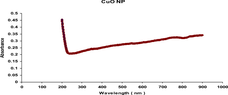
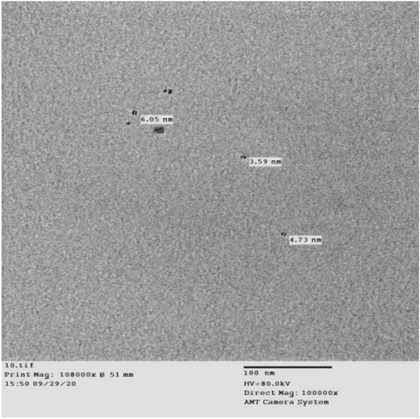
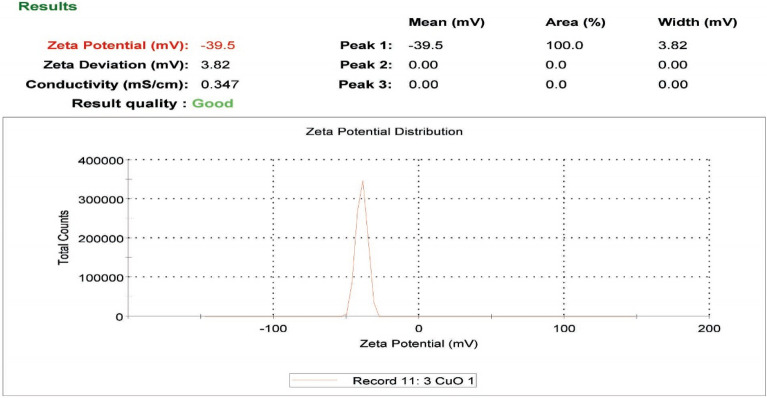
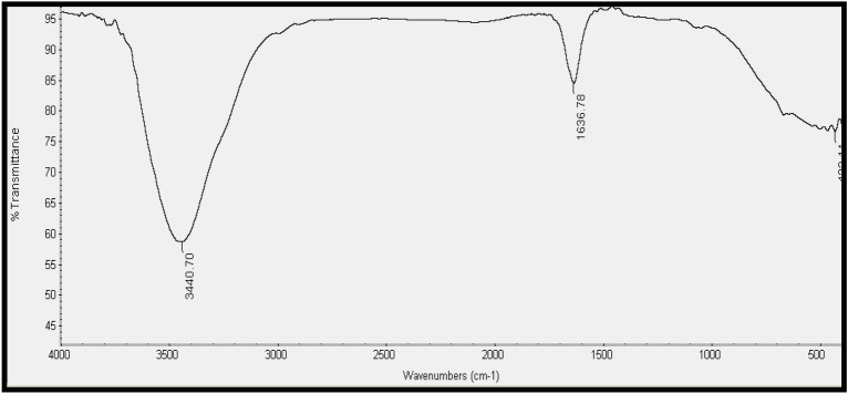
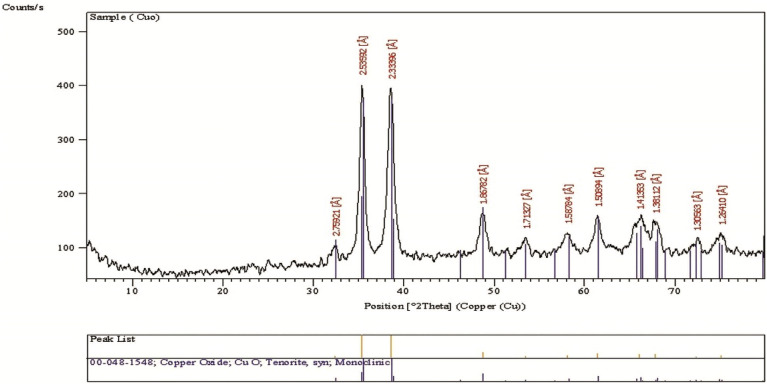
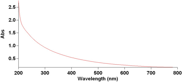
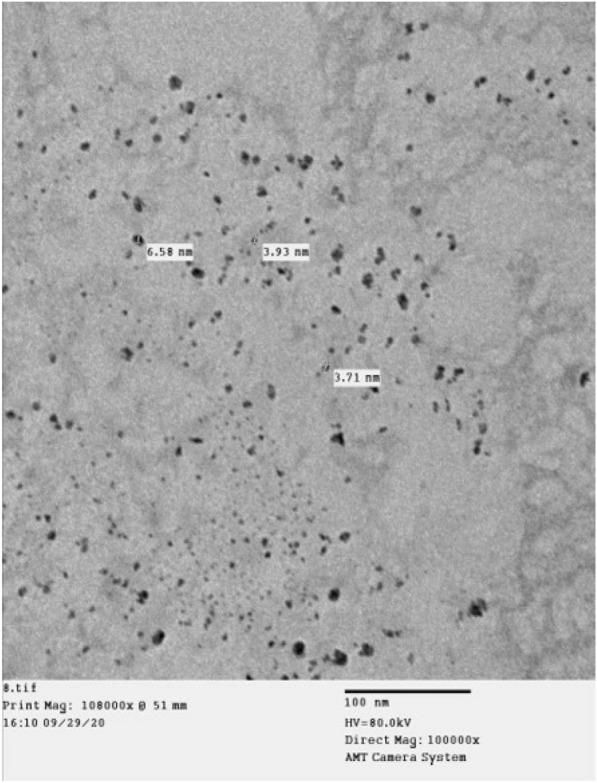
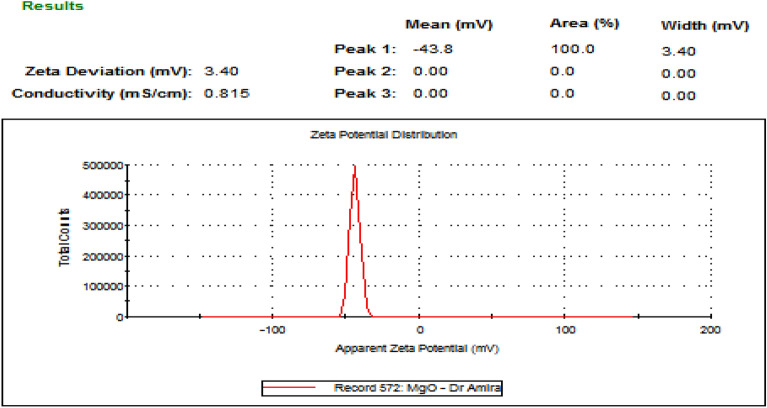
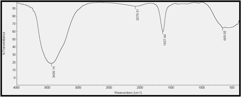
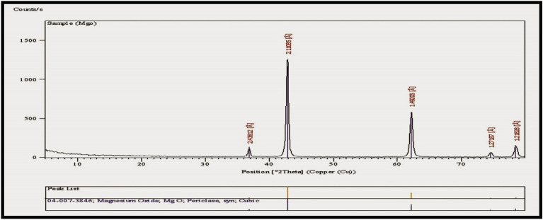
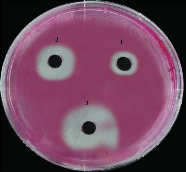
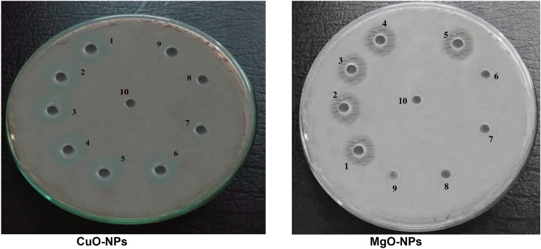
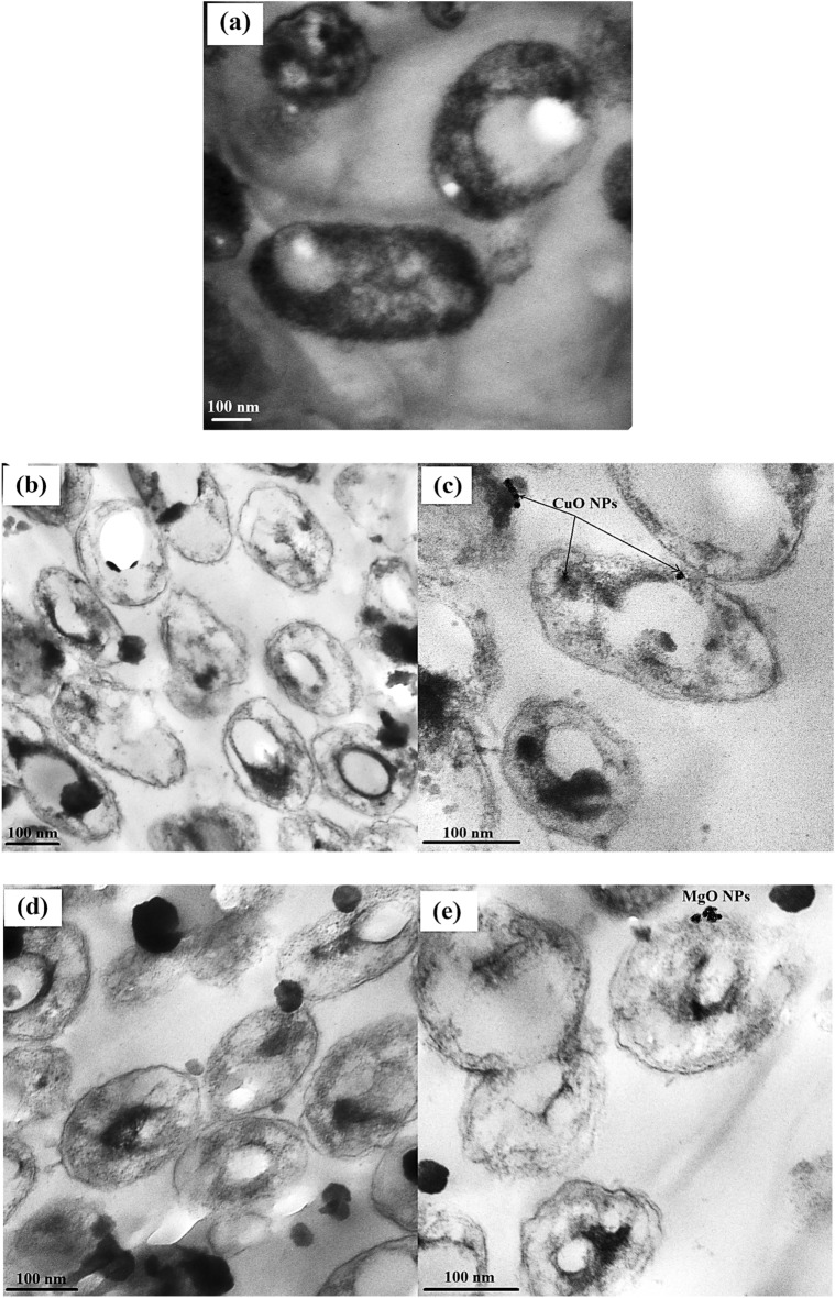
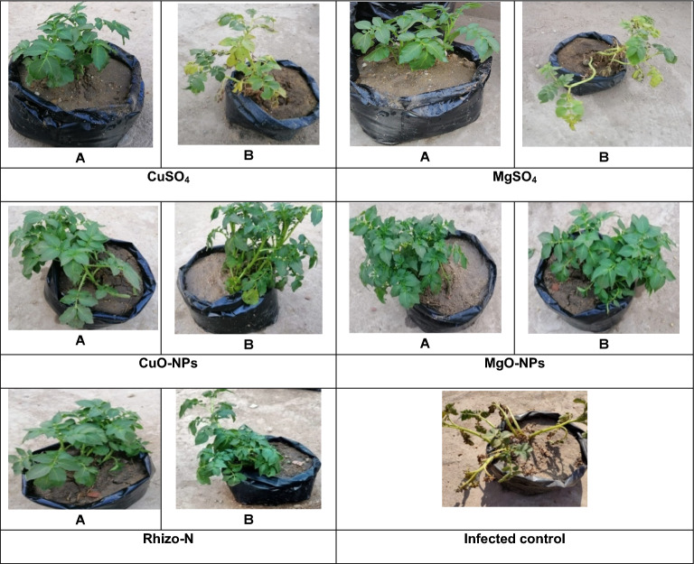
Results: CuO and MgO metal oxide nanoparticles were synthesized via a simple chemical process. The average particle size, morphology, and structure of the nanoparticles were characterized using UV-visible spectroscopy, transmission electron microscopy (TEM), zeta potential analysis, X-ray diffraction (XRD), and Fourier transform infrared (FTIR) spectroscopy. The growth of R. solanacearum was strongly inhibited by CuO and MgO NPs at a concentration of 3 mg/mL, resulting in zones of inhibition (ZOI) of 19.3 mm and 17 mm, respectively. The minimum inhibitory concentration (MIC) and minimum bactericidal concentration (MBC) of CuO-NPs and MgO-NPs were 0.5, 0.6, and 0.6, 0.75 mg/mL, respectively. When applied in vivo through seed dressing and tuber soaking at their respective MIC concentrations, CuO-NPs and MgO-NPs significantly reduced the incidence of brown rot disease to 71.2% and 69.4%, respectively, compared to 43.0% and 39.5% in bulk CuSO4 and bulk MgSO4 treatments, respectively. Furthermore, CuO-NPs and MgO-NPs significantly increased the yield, total chlorophyll content, and enzyme efficiency of potato plants compared with the infected control plants. TEM revealed that the bacterial cytomembrane was severely damaged by nanomechanical forces after interaction with CuO-NPs and MgO-NPs, as evidenced by lipid peroxidation and ultrastructural investigations.
Conclusion: The results of this study suggest that CuO-NPs and MgO-NPs can be used as intelligent agents to manage plant pathogens in agriculture. The use of metal oxide nanoparticles could provide a risk-free alternative for treating plant diseases, which are currently one of the biggest challenges faced by the potato industry in Egypt. The significant increase in yield, photosynthetic pigments, enzymatic activity, and total phenol-promoted resistance to R. solanacearum in potato plants treated with CuO-NPs and MgO-NPs compared to infected control plants highlights the potential benefits for the potato industry in Egypt. Further investigations are needed to explore using metal oxide nanoparticles for treating other plant diseases.
Keywords: Brown rot disease; Chlorophyll; CuO-NPs; Lipid peroxidation; MgO-NPs; Potato; R. solanacearum.
© 2023. The Author(s).
Conflict of interest statement
The authors declare no competing interests.
Figures
References
-
- Agredo–Trochez YA, Molano-Cabezas AC, Arciniegas-Grijalba PA, Rodríguez-Páez JE. Nanoparticles of magnesium oxyhydroxide and copper oxide: synthesis and evaluation of their in vitro fungicidal activity on the fungus Omphalia sp. Inorg Chem Comm. 2022;146:110085. doi: 10.1016/j.inoche.2022.110085. - DOI
-
- Ahmed W, Yang J, Tan Y, Munir S, Liu Q, Zhang J, Ji G, Zhao Z. Ralstonia solanacearum, a deadly pathogen: revisiting the bacterial wilt biocontrol practices in tobacco and other Solanaceae. Rhizosphere. 2022;22:100479. doi: 10.1016/j.rhisph.2022.100479. - DOI
-
- Baqer AA, Matori KA, Al-Hada NM, Kamari HM, Shaari AH, Saion E, Chyi JLY. Copper oxide nanoparticles synthesized by a heat treatment approach with structural, morphological and optical characteristics. J Mater Sci: Mater Electron. 2018;29:1025–1033. doi: 10.1007/s10854-017-8002-3. - DOI
LinkOut - more resources
Full Text Sources
