. 2023 Oct;45(5):2951-2965.
doi: 10.1007/s11357-023-00845-y. Epub 2023 Jul 17.

Targeting mitochondria in the aged cerebral vasculature with SS-31, a proteomic study of brain microvessels

Affiliations

Targeting mitochondria in the aged cerebral vasculature with SS-31, a proteomic study of brain microvessels

Abigail Seman et al. Geroscience. 2023 Oct.

Abstract

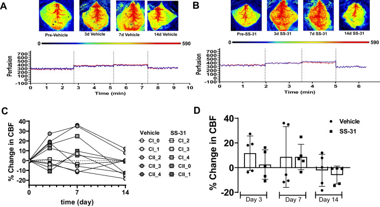
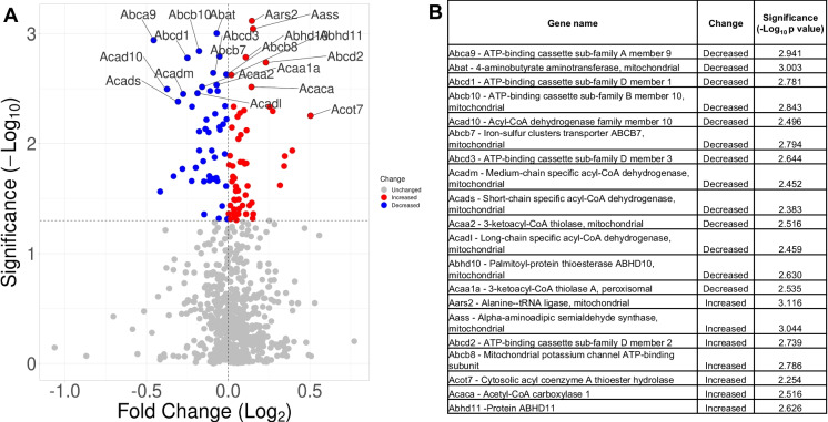
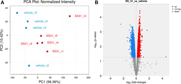
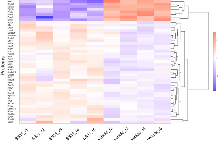
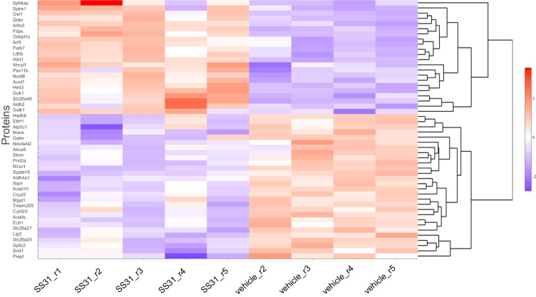
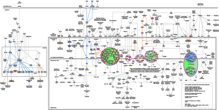
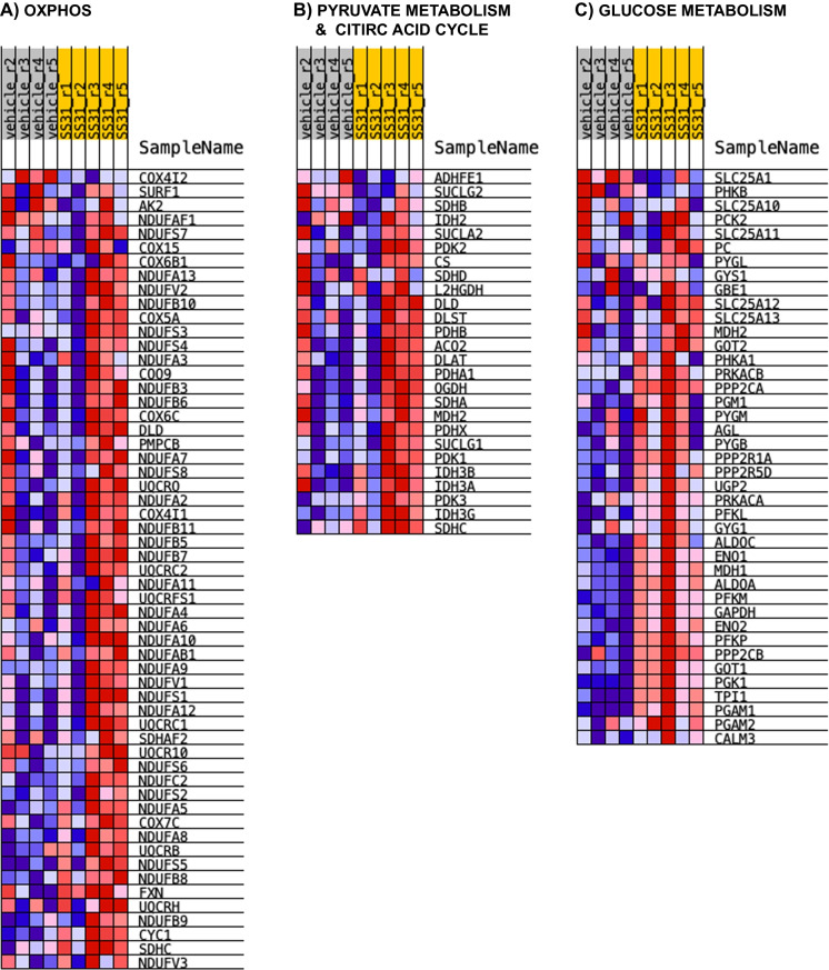
Cognitive impairment and dementias during aging such as Alzheimer's disease are linked to functional decline and structural alterations of the brain microvasculature. Although mechanisms leading to microvascular changes during aging are not clear, loss of mitochondria, and reduced efficiency of remaining mitochondria appear to play a major role. Pharmacological agents, such as SS-31, which target mitochondria have been shown to be effective during aging and diseases; however, the benefit to mitochondrial- and non-mitochondrial proteins in the brain microvasculature has not been examined. We tested whether attenuation of aging-associated changes in the brain microvascular proteome via targeting mitochondria represents a therapeutic option for the aging brain. We used aged male (> 18 months) C57Bl6/J mice treated with a mitochondria-targeted tetrapeptide, SS-31, or vehicle saline. Cerebral blood flow (CBF) was determined using laser speckle imaging during a 2-week treatment period. Then, isolated cortical microvessels (MVs) composed of end arterioles, capillaries, and venules were used for Orbitrap Eclipse Tribrid mass spectrometry. CBF was similar among the groups, whereas bioinformatic analysis revealed substantial differences in protein abundance of cortical MVs between SS-31 and vehicle. We identified 6267 proteins, of which 12% were mitochondria-associated. Of this 12%, 107 were significantly differentially expressed and were associated with oxidative phosphorylation, metabolism, the antioxidant defense system, or mitochondrial dynamics. Administration of SS-31 affected many non-mitochondrial proteins. Our findings suggest that mitochondria in the microvasculature represent a therapeutic target in the aging brain, and widespread changes in the proteome may underlie the rejuvenating actions of SS-31 in aging.

Keywords: Aging; Brain microvasculature; Mitochondria; Proteomics.


Conflict of interest statement

The authors declare no competing interests.

Figures

Fig. 1
Fig. 1
SS-31 does not affect baseline CBF. AB Representative images and CBF recordings from vehicle (saline) and SS-31-treated animals, respectively. The perfusion intensity scale, below the images, indicates the level of brain perfusion. The deep blue color on the perfusion intensity scale denotes “0” or no perfusion, whereas red color corresponds to “590” or well-perfused brain areas. Blue and red lines of CBF recordings are indicative of CBF on the two hemispheres. C Shows the percent changes in CBF of individual animals, whereas panel D represents the summary of CBF changes in the groups, stratified by measurement days. Data expressed as mean ± SD; n = 5 mice in the SS-31 group and n = 5 mice in the vehicle-treated group
Fig. 2
Fig. 2
Effects of SS-31 on mitochondrial/mitochondria-related protein abundance in aged brain MVs. A 107 proteins, associated with mitochondria, were significantly differentially expressed in response to SS-31 treatment when compared with vehicle. B Table shows the top 20 significantly differentially expressed mitochondria-associated proteins, labeled in panel A using the mitochondrial/mitochondria-related data set
Fig. 3
Fig. 3
SS-31 treatment affects protein abundance of aged MVs. A Principal component analysis (PCA) plot shows protein expression profiles of brain MVs from SS-31 (red color) and vehicle (blue color)-treated aged mice. PC1 and PC2: Principal components 1 and 2, respectively. B Volcano plot shows significantly differentially expressed proteins between the SS-31 and vehicle groups in the global proteome of aged MVs. Red color indicates protein upregulation, blue color corresponds to protein downregulation, whereas gray denotes not significantly differentially expressed proteins in response to SS-31 treatments in isolated brain microvessels
Fig. 4
Fig. 4
Heatmap of top 50 proteins in aged MVs. Heatmap shows the top 50 significantly differentially expressed proteins in aged MVs between the SS-31 and vehicle groups. n = 5 mice in the SS-31 group and n = 4 mice in the vehicle-treated group (1 vehicle was excluded from analysis)
Fig. 5
Fig. 5
Heatmap of top 50 mitochondria-associated proteins in aged MVs. Heatmap shows the top 50 significantly differentially expressed mitochondria associated proteins in aged MVs between the SS-31 and vehicle groups. n = 5 mice in the SS-31 group and n = 4 mice in the vehicle treated group (1 vehicle was excluded from analysis)
Fig. 6
Fig. 6
Mitochondrial dysfunction pathway. QIAGEN Ingenuity Pathway Analysis shows differentially expressed genes in our mitochondria only data set from SS-31 and vehicle-treated isolated brain microvessels from male, aged C57Bl/6J mice that are associated with mitochondrial dysfunction and oxidative phosphorylation canonical pathways
Fig. 7
Fig. 7
Gene Enrichment Set Analysis (GESA) heatmaps. SS-31 resulted in the enrichment of several proteins associated with A biological processes (GO_ OXIDATIVE_PHOSPHORYLATION) and B pyruvate metabolism and citric acid cycle (Reactome) within the mitochondrial data set, and in those identified in C glucose metabolism within the global data set

References

    1. Ungvari Z, Tarantini S, Kiss T, Wren JD, Giles CB, Griffin CT, et al. Endothelial dysfunction and angiogenesis impairment in the ageing vasculature. Nat Rev Cardiol. 2018;15(9):555–65. doi: 10.1038/s41569-018-0030-z. - DOI - PMC - PubMed
    1. Ungvari Z, Tarantini S, Donato AJ, Galvan V, Csiszar A. Mechanisms of vascular aging. Circ Res. 2018;123(7):849–67. doi: 10.1161/CIRCRESAHA.118.311378. - DOI - PMC - PubMed
    1. Nyul-Toth A, Tarantini S, DelFavero J, Yan F, Balasubramanian P, Yabluchanskiy A, et al. Demonstration of age-related blood-brain barrier disruption and cerebromicrovascular rarefaction in mice by longitudinal intravital two-photon microscopy and optical coherence tomography. Am J Physiol Heart Circ Physiol. 2021;320(4):H1370–H92. doi: 10.1152/ajpheart.00709.2020. - DOI - PMC - PubMed
    1. Brown WR, Thore CR. Review: cerebral microvascular pathology in ageing and neurodegeneration. Neuropathol Appl Neurobiol. 2011;37(1):56–74. doi: 10.1111/j.1365-2990.2010.01139.x. - DOI - PMC - PubMed
    1. Tracy EP, Hughes W, Beare JE, Rowe G, Beyer A, LeBlanc AJ. Aging-induced impairment of vascular function: mitochondrial redox contributions and physiological/clinical implications. Antioxid Redox Signal. 2021;35(12):974–1015. doi: 10.1089/ars.2021.0031. - DOI - PMC - PubMed

Publication types