. 2023 Aug 1;62(15):2269-2279.
doi: 10.1021/acs.biochem.3c00155. Epub 2023 Jul 17.

Post- and Pre-Radiolabeling Assays for anti Thymidine Cyclobutane Dimers as Intrinsic Photoprobes of Various Types of G-Quadruplexes, Reverse Hoogsteen Hairpins, and Other Non-B DNA Structures

Affiliations

Post- and Pre-Radiolabeling Assays for anti Thymidine Cyclobutane Dimers as Intrinsic Photoprobes of Various Types of G-Quadruplexes, Reverse Hoogsteen Hairpins, and Other Non-B DNA Structures

Natalia E Gutierrez-Bayona et al. Biochemistry. .

Abstract

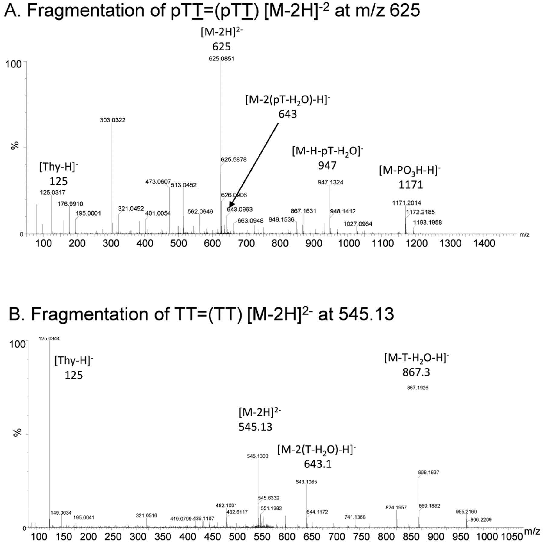
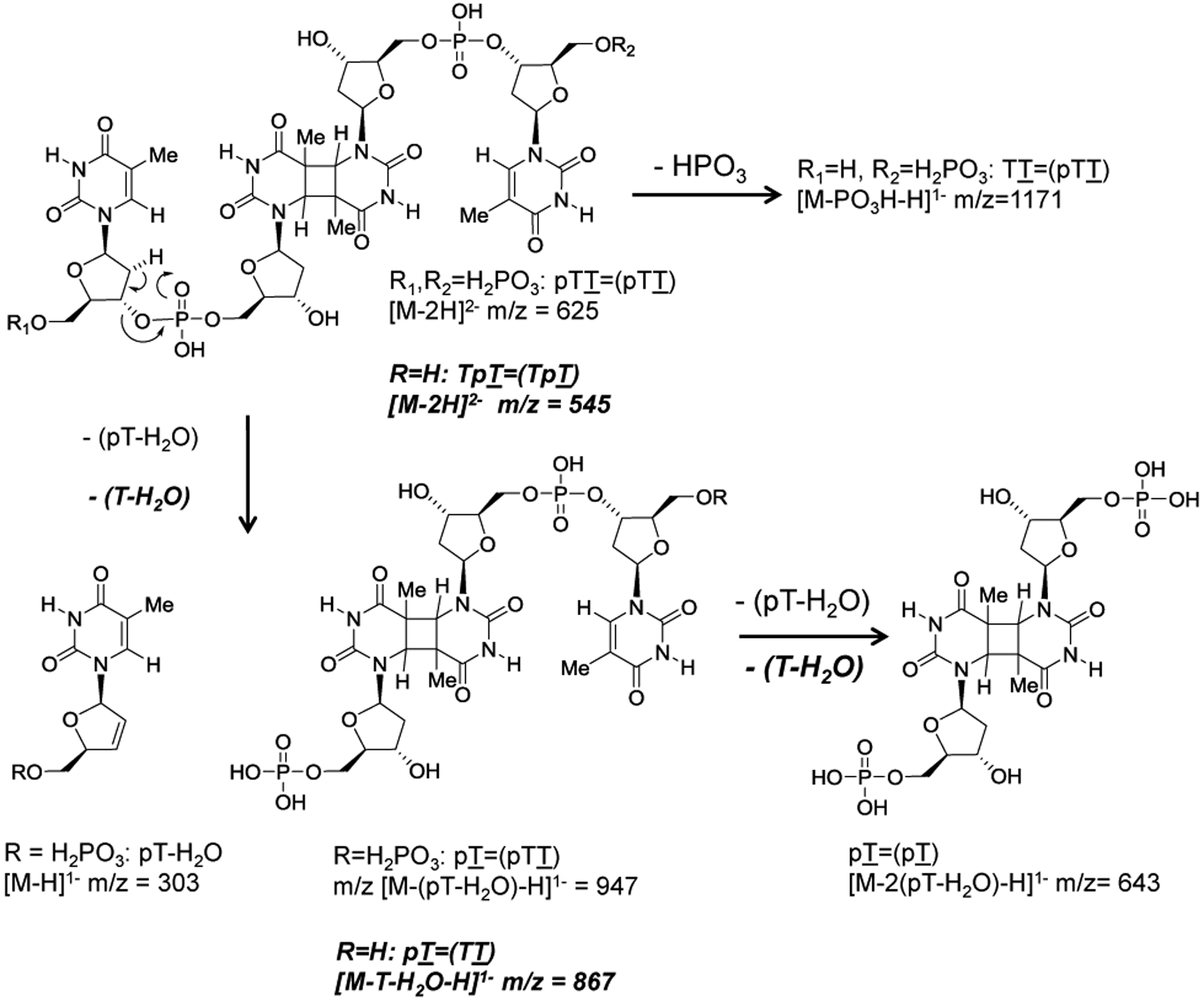
G-quadruplexes are thought to play an important role in gene regulation and telomere maintenance, but developing probes for their presence and location is challenging due to their transitory and highly dynamic nature. The majority of probes for G-quadruplexes have relied on antibody or small-molecule binding agents, many of which can also alter the dynamics and relative populations of G-quadruplexes. Recently, it was discovered that ultraviolet B (UVB) irradiation of human telomeric DNA and various G-quadruplex forming sequences found in human promoters, as well as reverse Hoogsteen hairpins, produces a unique class of non-adjacent anti cyclobutane pyrimidine dimers (CPDs). Therefore, one can envision using a pulse of UVB light to irreversibly trap these non-B DNA structures via anti CPD formation without perturbing their dynamics, after which the anti CPDs can be identified and mapped. As a first step toward this goal, we report radioactive post- and pre-labeling assays for the detection of non-adjacent CPDs and illustrate their use in detecting trans,anti T=(T) CPD formation in a human telomeric DNA sequence. Both assays make use of snake venom phosphodiesterase (SVP) to degrade the trans,anti T=(T) CPD-containing DNA to the tetranucleotide pTT=(pTT) corresponding to CPD formation between the underlined T's of two separate dinucleotides while degrading the adjacent syn TT CPDs to the trinucleotide pGT=T. In the post-labeling assay, calf intestinal phosphodiesterase is used to dephosphorylate the tetranucleotides, which are then rephosphorylated with kinase and [32P]-ATP to produce radiolabeled mono- and diphosphorylated tetranucleotides. The tetranucleotides are confirmed to be non-adjacent CPDs by 254 nm photoreversion to the dinucleotide p*TT. In the pre-labeling assay, radiolabeled phosphates are introduced into non-adjacent CPD-forming sites by ligation prior to irradiation, thereby eliminating the dephosphorylation and rephosphorylation steps. The assays are also demonstrated to detect the stereoisomeric cis,anti T=(T) CPD.


Conflict of interest statement

All authors declare no competing financial interest.

Figures

Figure 1.
Figure 1.
Idea of using structure-specific photoproducts to detect and map folded DNA structures such as basket G-quadruplexes. A) Structure of adjacent cis,syn and non-adjacent cis and trans,anti T=(T) CPDs where the parentheses denotes non-adjacent nucleotides. B) Whereas UVB irradiation of B DNA produces adjacent cis,syn T=T CPDs, UVB irradiation of non-B DNA folded structures such as the basket G-quadruplex form of human telomeric DNA produces unique non-adjacent anti T=(T) CPDs.
Figure 2.
Figure 2.
Post-labeling assay schemes for CPDs. A) DNA containing an adjacent cis,syn T=T CPD is degraded to a trinucleotide with snake venom phosphodiesterase (SVP) and then dephosphorylated with calf intestinal phosphatase (CIP) followed by radiolabeling with [γ-32P]-ATP and kinase to give a phosphorylated trinucleotide product. Irradiation with 254 nm light reverts the CPD to the canonical bases. B) DNA containing a non-adjacent anti T=(T) CPD is degraded to a tetranucleotide with snake venom phosphodiesterase (SVP) and after dephosphorylation with calf intestinal phosphatase (CIP) and radiolabeling with [γ-32P]-ATP and kinase would give mono and diphosphorylated tetranucleotide products. Irradiation with 254 nm light would revert the tetranucleotide products to two dinucleotides. Treatment of the tetra- or dinucleotide products with NP1 would release the 5’-terminal radiolabeled nucleotide. All radiolabeled products are distinguishable by high resolution polyacrylamide gel electrophoresis.
Figure 3.
Figure 3.
HPLC analysis and purification of the intermediates in the post-labeling assay of Tel26 DNA irradiated with UVB light. A) UVB irradiated Tel26 digested with snake venom phosphodiesterase (SVP) for 24 h to give trans,anti pTT=(pTT). B) Dephosphorylation of trans,anti pTT=(pTT) with calf intestinal phosphodiesterase (CIP) to give TT=(TT). C) Rephosphorylation of TT=(TT) from B with polynucleotidyl kinase (PNK) and ATP.
Figure 4.
Figure 4.
ESI-MS/MS characterization of the intermediates in the post-labeling assay of irradiated Tel26 DNA. A) MS/MS of the [M - 2H]2- ion (m/z 624.11) of trans,anti pTT=(pTT). b) MS/MS of the [M - 2H]2- ion (m/z 545.13) of the dephosphorylated trans,anti TT=(TT).
Figure 5.
Figure 5.
MS/MS fragmentation pathways of the trans,anti T=(T) CPD-containing products of Figure 4. Pathways for pTT=(pTT) are shown in normal font, whereas pathways for TT=(TT) are shown in bold italic font.
Figure 6.
Figure 6.
Post-labeling assay carried out on UVB irradiated Tel26 in the absence or presence of competing plasmid DNA. Lanes 2, 3 and 5 are authentic standards prepared by radiolabeling TT and HPLC purified GT=T and TT=(TT). 254 nm light was used to photorevert CPD-containing products. Lane headings for the post-labeling reactions refer to the ratio of the base pair concentrations of Tel26 to plasmid DNA. The portion of the gel to the right is shown with a reduced dynamic range to make the photoreversed dinucleotide bands more apparent. An asterisk indicates a radiolabeled phosphate.
Figure 7.
Figure 7.
Preparation and use of internally labeled Tel26 for studies of anti T=(T) CPD formation. A) ODNs (written 5’−3’) used to prepare Tel26 internally labeled in loop 1 and loop 2 where the T’s that undergo trans,anti T=(T) CPD formation are in bold. The 30-mer serves as a ligation scaffold. B) Degradation products the non-adjacent trans,anti T=(T) CPD formed between loop 1 and loop 3 of Tel26 internally labeled at loop 1. The relative mobility of the enzymatic degradation products is compared to the mass to charge ratio assuming that all phosphate groups are fully ionized. An asterisk indicates a radiolabeled phosphate.
Figure 8.
Figure 8.
Analysis of the enzymatic degradation products of post- and pre-labeled Tel26 by denaturing polyacrylamide gel electrophoresis. Lanes 1–3: authentic products obtained by end-labeling commercial ODNs. Lanes 4–7: post-labeling assay carried out on UVB irradiated Tel26 followed by dephosphorylation with CIP, with and without CPD photoreversion with 254 nm light as indicated. Lanes 8–13: SVP treatment of UVB irradiated Tel26 that was internally pre-labeled in loop 1 followed by CIP or CIP and NP1, with and without CPD photoreversion with 254 nm light, all as indicated. Lanes 14–18: SVP treatment of UVB irradiated Tel26 that was internally pre-labeled in loop 2 followed by CIP with and without CPD photoreversion with 254 nm light as indicated. Post treatments with CIP (Post CIP) were for either 2.5 min or 30 min as indicated by a or b in the lane headings. Figure prepared from two separate gels carried out under identical conditions as indicated by the vertical line. An asterisk indicates a radiolabeled phosphate.
Figure 9.
Figure 9.
Analysis of the enzymatic degradation products of pre- and post-labeled 14-mer containing a cis,anti T=(T) CPD. A) Schematic for the enzymatic reactions carried out on the pre- and post-labeled UVB irradiated 14-mer where the T’s undergoing anti CPD formation are in bold. B) Denaturing PAGE of the enzymatic reaction products. Lanes 5–12: the 5’-pre-labeled UVB irradiated 14-mer was digested with SVP and then dephosphorylated with CIP, where a, b and c refer to the times of 0-, 5- and 10-minutes following addition of CIP, with and without photoreversion by 254 nm light as indicated. Lanes 13–20: The UVB irradiated 14-mer was degraded by SVP and then labeled with PNK and [γ-32P]-ATP, after which it was incubated for increasing time with CIP as described for the pre-labeled experiment, with and without photoreversion with 254 nm light as indicated. An asterisk indicates a radiolabeled phosphate.

Similar articles

References

    1. Sinden RR Slipped strand DNA structures. Front. Biosci 2007, 12, 4788. - PubMed
    1. Saini N, Zhang Y, Usdin K, and Lobachev KS When secondary comes first - the importance of non-canonical DNA structures. Biochimie 2013, 95, 117–123. - PMC - PubMed
    1. Thys RG, Lehman CE, Pierce LC, and Wang YH DNA secondary structure at chromosomal fragile sites in human disease. Curr. Genomics 2015, 16, 60–70. - PMC - PubMed
    1. Sullivan ED, Longley MJ, and Copeland WC Polymerase γ efficiently replicates through many natural template barriers but stalls at the HSP1 quadruplex. J. Biol. Chem 2020, 295, 17802–17815. - PMC - PubMed
    1. Bochman ML, Paeschke K, and Zakian VA DNA secondary structures: stability and function of G-quadruplex structures. Nat. Rev. Genet 2012, 13, 770–780. - PMC - PubMed

Publication types