Review
. 2023 Nov;221(5):633-648.
doi: 10.2214/AJR.23.29003. Epub 2023 Jun 7.

Staging of Cervical Cancer: A Practical Approach Using MRI and FDG PET

Affiliations
Review

Staging of Cervical Cancer: A Practical Approach Using MRI and FDG PET

Yulia Lakhman et al. AJR Am J Roentgenol. 2023 Nov.

Abstract

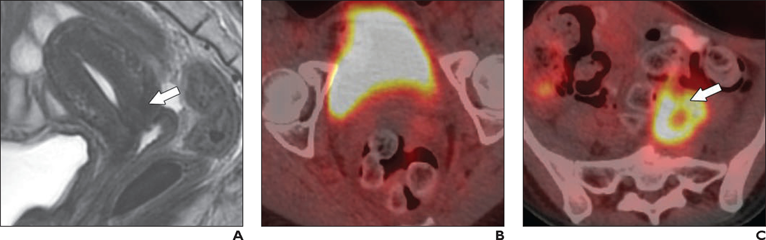
This review provides a practical approach to the imaging evaluation of patients with cervical cancer (CC), from initial diagnosis to restaging of recurrence, focusing on MRI and FDG PET. The primary updates to the International Federation of Gynecology and Obstetrics (FIGO) CC staging system, as well as these updates' relevance to clinical management, are discussed. The recent literature investigating the role of MRI and FDG PET in CC staging and image-guided brachytherapy is summarized. The utility of MRI and FDG PET in response assessment and posttreatment surveillance is described. Important findings on MRI and FDG PET that interpreting radiologists should recognize and report are illustrated. The essential elements of structured reports during various phases of CC management are outlined. Special considerations, including the role of imaging in patients desiring fertility-sparing management, differentiation of CC and endometrial cancer, and unusual CC histologies, are also described. Finally, future research directions including PET/MRI, novel PET tracers, and artificial intelligence applications are highlighted.

Keywords: MRI; PET; cervical cancer; gynecologic cancer; imaging.


Figures

Fig. 1—
Fig. 1—
Illustrations show cervical cancer stages according to International Federation of Gynecology and Obstetrics (FIGO) staging system [6, 7]. A, Cervix-confined invasive carcinoma is deemed FIGO stage I, which is divided into stage IA and stage IB. All stage IA tumors (not shown) are microscopic with maximum depth of stromal invasion of 5 mm or less. If stromal invasion is 3 mm or less, tumor is deemed stage IA1; if stromal invasion is from more than 3 mm to 5 mm, tumor is deemed stage IA2. All stage IB tumors (yellow) show stromal invasion of more than 5 mm in depth. Stage IB tumors that are 2 cm or smaller in greatest dimension are deemed stage IB1; more than 2 cm to 4 cm, stage IB2; and more than 4 cm, IB3. B, In FIGO stage II, tumor has spread beyond cervix but has not spread into lower third of vagina or into pelvic sidewall. Stage IIA tumors (yellow, upper panel) involve upper two-thirds of vagina but not parametria, with stage IIA1 tumors being 4 cm or less in greatest dimension and stage IIA2 tumors measuring more than 4 cm in greatest dimension. Stage IIB tumors (yellow, lower panel) show parametrial invasion. C, FIGO stage III tumors show spread beyond what is seen with stage II. Tumor (yellow, left panel) spread to lower third of vagina is stage IIIA. Stage IIIB involves one or more of: pelvic sidewall extension (yellow, middle panel), hydronephrosis, or nonfunctioning kidney. Tumors metastasizing to lymph nodes (LNs) (yellow, right panel) are stage IIIC: tumors metastasizing to pelvic LNs only are considered stage IIIC1 and tumors metastasizing to paraaortic LNs are considered stage IIIC2. D, FIGO stage IV tumors involve bladder mucosa or rectum (proven by biopsy) or spread beyond true pelvis. In stage IVA, tumor (yellow, left panel) invades adjacent organs in pelvis, although bullous edema of bladder or rectum only is not enough to assign stage IVA. Stage IVB tumors show distant metastases, including spread to LNs beyond pelvic and paraaortic regions (yellow, right panel).
Fig. 2—
Fig. 2—
Diagram illustrates how International Federation of Gynecology and Obstetrics stage informs management of cervical cancer. One asterisk indicates that paraaortic region may be included in radiation treatment if there is suspicion for common iliac lymph node (LN) metastases; two asterisks indicate that paraaortic lymphadenectomy may be performed if common iliac and/or paraaortic LN metastases are suspected on basis of FDG PET findings.
Fig. 3—
Fig. 3—
Relevant anatomy of cervix and surrounding structures. A, Illustration shows sagittal view of uterus, cervix, vagina, and urinary bladder. Location of internal os (dotted line) is seen as narrowing between endocervical canal and endometrial cavity. Yellow dashed line illustrates orientation for oblique axial plane. Horizontal line at level of bladder neck approximately divides vagina into upper two-thirds and lower one-third. B, Illustration shows that location of internal os in oblique axial plane is marked by entrance of uterine vessels (red and blue). Parametrium is connective tissues lateral to cervix.
Fig. 4—
Fig. 4—
35-year-old woman with newly diagnosed invasive squamous cell cervical carcinoma and desire for fertility preservation. A and B, Sagittal (A) and oblique axial (B) T2-weighted images show 1.2-cm exophytic intermediatesignal cervix-confined tumor (arrows) consistent with International Federation of Gynecology and Obstetrics stage IB1 disease. Tumor is located away from internal cervical os (arrowhead, A). C, Axial fused image of T2-weighted image and DWI shows restricted diffusion within tumor (arrow). D, Sagittal T2-weighted image obtained after radical trachelectomy shows anastomosis (arrows) between remaining uterus and vagina.
Fig. 5—
Fig. 5—
Features on T2-weighted MRI that are useful for determining presence of parametrial invasion of cervical cancer (yellow). A, Illustration shows partial- or full-thickness cervical stromal invasion, which is not sufficient to diagnose parametrial invasion. B, Illustrations show parametrial invasion, which is indicated by full-thickness stromal invasion and either nodular (left) or spiculated (right) interface with parametrium without or with parametrial vessel (red and blue, right panel) encasement.
Fig. 6—
Fig. 6—
Evaluation of cervical stroma invasion and parametrial invasion (PMI). A, 46-year-old woman with invasive squamous cell carcinoma. Oblique axial T2-weighted image shows 3.3-cm tumor with full-thickness cervical stromal invasion but smooth outer contour (arrow). Surgical pathology revealed near-full-thickness (16 of 17 mm) cervical stromal invasion and lack of PMI. Findings are consistent with International Federation of Gynecology and Obstetrics (FIGO) stage IB2 disease. B and C, 52-year-old woman with invasive squamous cell carcinoma. Oblique axial T2-weighted image (B) and axial fused image (T2-weighted imaging and DWI) (C) show full-thickness cervical stromal invasion and nodular outer contour (arrow, B) indicative of PMI. Findings are consistent with FIGO stage IIB disease.
Fig. 7—
Fig. 7—
64-year-old woman with invasive squamous cell carcinoma. A and B, Sagittal (A) and oblique axial (B) T2-weighted images show large ill-defined cervical tumor (arrow) with involvement of lower one-third of vagina and urinary bladder lumen. Few areas of bullous edema are present within mucosa. In A, arrow shows tumor. Arrowhead in B shows bullous edema. Findings are consistent with International Federation of Gynecology and Obstetrics stage IVA disease.
Fig. 8—
Fig. 8—
28-year-old woman with invasive squamous cell carcinoma of cervix. A, Sagittal T2-weighted image shows large exophytic intermediate-signal tumor (T). B, Oblique axial T2-weighted image shows enlarged external bilateral iliac lymph nodes (LNs) (arrows) with signal intensity similar to that of primary cervical tumor. C, Oblique axial high-b-value DW image shows enlarged external bilateral iliac LNs (arrows) with high signal intensity. 28-year-old woman with invasive squamous cell carcinoma of cervix. D and E, Axial fused images from FDG PET/CT show FDG-avid primary tumor (T, D) and bilateral external iliac LNs (arrows, D) and left common iliac LN (arrow, E). Findings are consistent with International Federation of Gynecology and Obstetrics stage IIIC disease.
Fig. 8—
Fig. 8—
28-year-old woman with invasive squamous cell carcinoma of cervix. A, Sagittal T2-weighted image shows large exophytic intermediate-signal tumor (T). B, Oblique axial T2-weighted image shows enlarged external bilateral iliac lymph nodes (LNs) (arrows) with signal intensity similar to that of primary cervical tumor. C, Oblique axial high-b-value DW image shows enlarged external bilateral iliac LNs (arrows) with high signal intensity. 28-year-old woman with invasive squamous cell carcinoma of cervix. D and E, Axial fused images from FDG PET/CT show FDG-avid primary tumor (T, D) and bilateral external iliac LNs (arrows, D) and left common iliac LN (arrow, E). Findings are consistent with International Federation of Gynecology and Obstetrics stage IIIC disease.
Fig. 9—
Fig. 9—
53-year-old woman with newly diagnosed adenocarcinoma of cervix. A, Axial T2-weighted image shows small right external iliac lymph node (LN) (arrow) measuring 6 mm in short axis. B, Coronal fused image from FDG PET/CT shows FDG avidity within LN (arrow). Findings are consistent with International Federation of Gynecology and Obstetrics stage IIIC1 disease.
Fig. 10—
Fig. 10—
42-year-old woman who presented with clear vaginal discharge and was subsequently diagnosed with International Federation of Gynecology and Obstetrics stage IB3 gastric-type adenocarcinoma of cervix. A, Sagittal T2-weighted image shows 6-cm infiltrative partly endophytic mass (arrow) replacing entire cervix; mass shows intermediate signal intensity and has internal microcysts (arrowhead). B, Sagittal fat-saturated contrast-enhanced T1-weighted image shows enhancement of mass (arrow).
Fig. 11—
Fig. 11—
28-year-old woman with invasive squamous cell carcinoma of cervix (same patient as in Fig. 8). A, Sagittal T2-weighted image obtained 5 months after definitive chemoradiotherapy shows reconstitution of hypointense cervical stroma (arrow), indicating absence of tumor. B, Axial fused image from FDG PET/CT shows resolution of FDG avidity within cervical tumor (compared with Fig. 8D). C, Axial fused image from FDG PET/CT shows increased FDG uptake in left common iliac lymph node (arrow) (compared with Fig. 8E); this finding is consistent with persistent metastatic disease.

Comment in

References

    1. Sung H, Ferlay J, Siegel RL, et al. Global cancer statistics 2020: GLOBOCAN estimates of incidence and mortality worldwide for 36 cancers in 185 countries. CA Cancer J Clin 2021; 71:209–249 - PubMed
    1. Stelzle D, Tanaka LF, Lee KK, et al. Estimates of the global burden of cervical cancer associated with HIV. Lancet Glob Health 2021; 9:e161–e169 - PMC - PubMed
    1. Lei J, Ploner A, Elfström KM, et al. HPV vaccination and the risk of invasive cervical cancer. N Engl J Med 2020; 383:1340–1348 - PubMed
    1. WHO website. Cervical cancer. www.who.int/news-room/fact-sheets/detail/cervical-cancer. Published Feb 22, 2022. Accessed Nov 7, 2022
    1. Parra-Herran C Cervix: WHO classification. PathologyOutlines.com website. www.pathologyoutlines.com/topic/cervixWHO.html. Last updated May 28, 2021. Accessed Nov 7, 2022

MeSH terms

Substances