Subepidermal calcified nodule (nodular calcinosis of Winer)
- PMID: 37460248
- PMCID: PMC10357789
- DOI: 10.1136/bcr-2023-255461
Subepidermal calcified nodule (nodular calcinosis of Winer)
Abstract
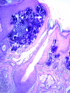
Subepidermal calcified nodule (SCN) is a clinical form of idiopathic calcinosis cutis, which commonly affects children, and presents as yellowish-white lesions involving the face. It is often misdiagnosed for other disorders like warts and molluscum contagiosum and treated by ablative procedures. In such a scenario, lack of histopathological examination makes it difficult to reach the correct diagnosis. We here report a case of SCN which was diagnosed after an excisional biopsy. Further, histopathological finding of dermal calcium deposits must prompt the clinician to rule out other disorders leading to calcinosis cutis, before labelling the case as SCN.
Keywords: Dermatology; General practice / family medicine; Pathology.
© BMJ Publishing Group Limited 2023. No commercial re-use. See rights and permissions. Published by BMJ.
Conflict of interest statement
Competing interests: None declared.
Figures


References
Publication types
MeSH terms
LinkOut - more resources
Full Text Sources
Medical
